. 2001 Sep;12(9):2742-55.
doi: 10.1091/mbc.12.9.2742.

Myosin Va bound to phagosomes binds to F-actin and delays microtubule-dependent motility

Affiliations
Free PMC article

Myosin Va bound to phagosomes binds to F-actin and delays microtubule-dependent motility

A Al-Haddad et al. Mol Biol Cell. 2001 Sep.
Free PMC article

Abstract

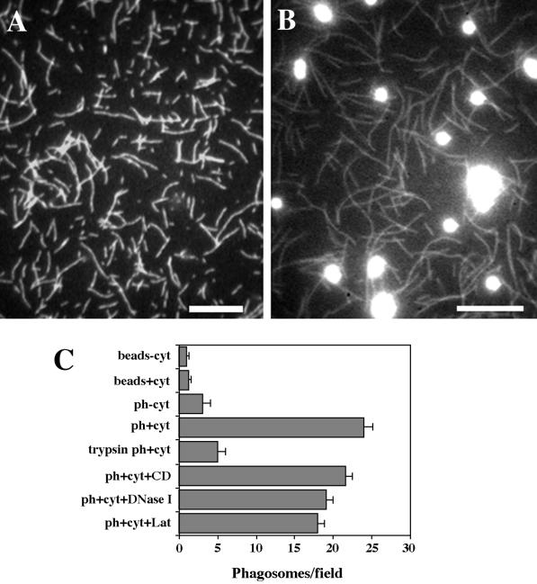
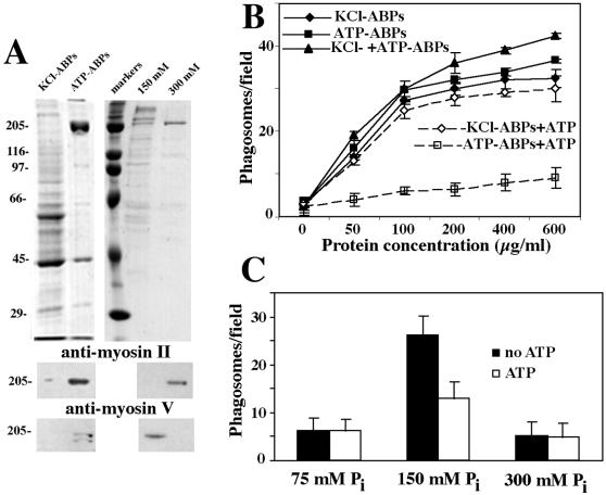
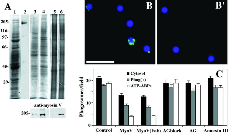
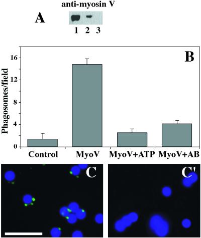
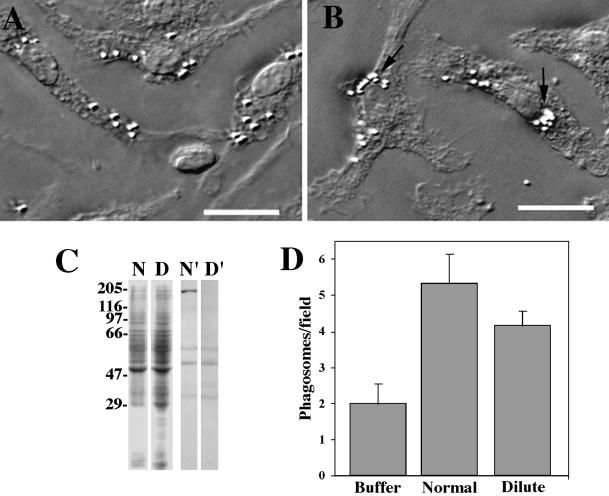
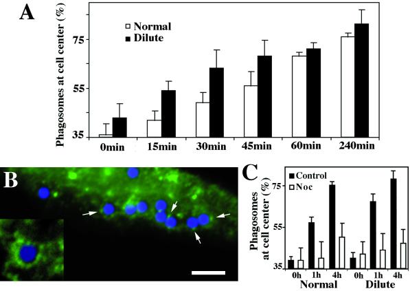
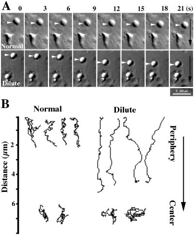
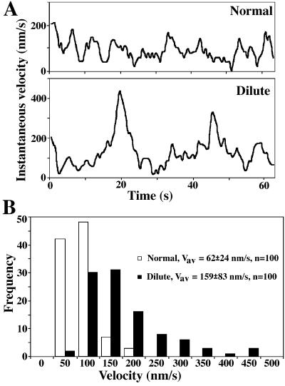
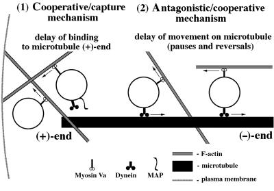
We established a light microscopy-based assay that reconstitutes the binding of phagosomes purified from mouse macrophages to preassembled F-actin in vitro. Both endogenous myosin Va from mouse macrophages and exogenous myosin Va from chicken brain stimulated the phagosome-F-actin interaction. Myosin Va association with phagosomes correlated with their ability to bind F-actin in an ATP-regulated manner and antibodies to myosin Va specifically blocked the ATP-sensitive phagosome binding to F-actin. The uptake and retrograde transport of phagosomes from the periphery to the center of cells in bone marrow macrophages was observed in both normal mice and mice homozygous for the dilute-lethal spontaneous mutation (myosin Va null). However, in dilute-lethal macrophages the accumulation of phagosomes in the perinuclear region occurred twofold faster than in normal macrophages. Motion analysis revealed saltatory phagosome movement with temporarily reversed direction in normal macrophages, whereas almost no reversals in direction were observed in dilute-lethal macrophages. These observations demonstrate that myosin Va mediates phagosome binding to F-actin, resulting in a delay in microtubule-dependent retrograde phagosome movement toward the cell center. We propose an "antagonistic/cooperative mechanism" to explain the saltatory phagosome movement toward the cell center in normal macrophages.


Figures

Figure 1
Figure 1
Reconstitution of salt-stripped phagosome binding to preassembled F-actin in vitro. (A and B) Fluorescence images of typical fields of the binding assay. (A) Rhodamine-phalloidin–labeled F-actin was absorbed on the coverslip surface. (B) Phagosomes were bound to F-actin in the presence of 1 mg/ml cytosol and unbound phagosomes were washed out. Bars, 10 μm. (C) Stimulation of phagosome binding to F-actin by cytosolic (cyt) factor(s). Binding to the F-actin lawn of uninternalized latex beads (beads−cyt; beads+cyt). Phagosome (ph) binding to F-actin in the presence and absence of 1 mg/ml cytosol (ph+cyt, ph−cyt). Binding of phagosomes treated with 100 μg/ml trypsin (trypsin ph+cyt) (see MATERIALS AND METHODS). Binding of phagosomes in the presence of 5 μM cytochalasin D (ph+cyt+CD), 15 μM DNase I (ph+cyt+DNase I), or 5 μM latrunculin (ph+cyt+Lat).
Figure 2
Figure 2
Involvement of ABPs in salt-stripped phagosome–F-actin interaction. (A) Analysis of ABP fractions by SDS-PAGE and immunoblotting. (Top) ABPs eluted from actin gel in the presence of 0.5 M KCl (KCl-ABPs), ABPs eluted from actin gel in the presence of 0.5 M KCl plus 2 mM ATP (ATP-ABPs), molecular mass markers (markers), and ATP-ABP fraction eluted from the HAP column in the presence of 150 mM Pi (150 mM) and in the presence of 300 mM Pi (300 mM) were run on 10% gel and stained with Coomassie blue. (Middle) Blot of the fractions described above probed with a monoclonal anti-myosin II heavy chain. (Bottom) Blot of corresponding fractions probed with a polyclonal antibody against the tail domain of chicken myosin V heavy chain. (B) Phagosome binding to F-actin in the presence of ABPs eluted from an actin gel without ATP (KCl-ABPs), with ATP (ATP-ABPs), and in the presence of both ABPs fractions (KCl+ATP-ABPs). ATP (2 mM) effects on phagosome binding in the presence of KCl-ABPs (KCl-ABPs+ATP), ATP-ABPs (ATP-ABPs+ATP). The protein concentrations for each ABP fraction are indicated. (C) Binding activity of ATP-ABPs after fractionation on the HAP column. Fractions were eluted from the column in the presence of 75, 150, and 300 mM Pi. For all fractions, the binding activity was tested in the presence (ATP) and absence (no ATP) of 2 mM ATP.
Figure 3
Figure 3
Myosin Va detection in phagosome preparations with associated F-actin–binding activity. (A) DIL2 antibody strongly reacted with myosin V in macrophage cytosol. (Lane 1) SDS-PAGE of cytosol (Coomassie staining). (Lane 2) Immunoblot probed with DIL2 antibody. Salt-stripped phagosomes were reisolated by flotation after incubation in the presence of cytosol at 20 mg/ml (lane 3) or 1 mg/ml (lane 4). The upper panel shows SDS-PAGE of phagosomes (Coomassie staining). The lower panel shows an immunoblot probed with DIL2 antibody. Reisolated (mock) salt-stripped phagosomes (lane 5) and phagosomes reisolated by flotation after incubation in the presence of ATP-ABPs (lane 6). The upper panel shows SDS-PAGE of phagosomes (silver staining). The lower panel shows an immunoblot probed with DIL2 antibody. (B and B′) Immunofluorescence labeling of isolated phagosomes with DIL2 antibody against myosin Va. (B) Phagosomes with F-actin–binding activity reisolated after preincubation with 1 mg/ml cytosol. Fluorescent patches were found associated with phagosomes. (B′) Phagosomes without F-actin–binding activity reisolated after preincubation with 20 mg/ml cytosol. No fluorescence signal was found associated with these phagosomes. Bar, 5 μm. (C) Inhibition of phagosome binding to F-actin with DIL2 antibody against myosin Va. Polyclonal DIL2 antibody (bars MyoV), Fab fragments [bars Myo(Fab)], and antigen-blocked DIL2 antibody (bars AGblock) were tested for effects on phagosome binding to F-actin when added directly to the binding reaction mixture in the presence of 2 mg/ml cytosol and salt-stripped phagosomes (cytosol), in the presence of reisolated phagosomes with F-actin-binding activity after preincubation with 2 mg/ml cytosol [Phag(+)], and in the presence of 0.1 mg/ml ATP-ABPs. Control, AG and annexin III show the effects on phagosome binding of no additions, DIL2 antigen alone, or control polyclonal antibody to annexin III (a protein normally associated with isolated phagosomes; Diakonova et al., 1997), respectively.
Figure 4
Figure 4
ATP-dependent interaction between phagosomes and F-actin in the presence of myosin Va isolated from chicken brain. (A) Immunoblot probed with polyclonal antibody against the head domain of chicken myosin Va (anti-myosin V). (Lane 1) Purified myosin Va from chicken brain. (Lane 2) Salt-stripped phagosomes reisolated by flotation after incubation in the presence of chicken myosin V at 50 μg/ml. (Lane 3) Reisolated (mock) salt-stripped phagosomes. (B) Phagosome binding to F-actin in the presence of chicken myosin Va. Mock phagosomes without binding activity (bar control). Stimulation of phagosome binding in the presence of 50 μg/ml chicken myosin Va (bar MyoV). Inhibition of phagosome binding to F-actin with 2 mM ATP (bar MyoV+ATP) and with polyclonal antibody against the head domain of chicken myosin Va (bar MyoV+AB). (C and C′) Immunofluorescence labeling of isolated phagosomes with polyclonal antibody against chicken myosin Va. (C) Phagosomes with F-actin–binding activity reisolated after preincubation with 50 μg/ml chicken myosin Va. (C′) Phagosomes without F-actin–binding activity reisolated by flotation after preincubation with buffer. Bar, 5 μm.
Figure 5
Figure 5
(A and B) Accumulation of the phagosomes in the perinuclear region of macrophages. Cells were loaded with latex beads for 15 min (A) and then chased for 4 h (B). Accumulation of phagosomes is seen at the cell centers (arrows). The same accumulation was observed in dilute-lethal macrophages (not shown). Bars, 10 μm. (C) Detection of myosin Va in normal and dilute-lethal macrophages. SDS-PAGE of lysates of normal (N) and dilute-lethal (D) macrophages; immunoblot of lysates of normal (N′) and dilute-lethal (D′) macrophages probed with DIL2 antibody. No myosin Va was detected in dilute-lethal macrophages. (D) Binding activity of phagosomes to F-actin in the absence of cytosol (buffer) and in the presence of 0.2 mg/ml cytosol isolated from normal macrophages (normal) or from dilute-lethal macrophages (dilute). The difference of stimulation between the two types of cytosol is ∼35%.
Figure 6
Figure 6
(A) Time course of phagosome accumulation near the cell center in normal and dilute-lethal macrophages based on three independent experiments. Cells were loaded for 15 min, washed for 15 min (0 time), and then chased for 15, 30, 45, 60, and 240 min. For each time point, 20 cells were analyzed (see MATERIALS AND METHODS). An increased rate of accumulation was found in dilute-lethal macrophages (see time points 15, 30, and 45 min). (B) Immunofluorescence labeling with DIL2 antibody of normal macrophages engaged in phagocytosis. Fluorescent patches were found associated with some phagosomes enclosing blue latex beads in normal macrophages (arrows), but not in dilute-lethal cells (not shown). Bar, 10 μm. (Inset) Twofold-higher magnification. (C) Time course of phagosome accumulation near the cell center in normal (normal) and dilute-lethal (dilute) macrophages in the presence of nocodazole. The cells were treated as in A and chased for 1 h and 4 h in media with 5 μM nocodazole (Noc).
Figure 7
Figure 7
Phagosome movement in normal and dilute-lethal macrophages. (A) Successive video frames (3-s intervals) of centripetal phagosome movements (arrowheads) in normal (normal) and dilute-lethal (dilute) macrophages. The black arrows indicate the direction of movement toward the nucleus. (B) Typical paths for phagosome movements that occurred over a 2-min period inward from the periphery of normal macrophages and dilute-lethal macrophages. In the lower part, typical paths in the perinuclear region (center) of normal and dilute-lethal macrophages are shown. The arrow indicates the directionality of movement toward the nucleus. Download movies at http://www.biologie.uni-rostock.de/abt/tierphys/kuznet/movies.htm.
Figure 8
Figure 8
Velocity of phagosome movement in the periphery of normal and dilute-lethal macrophages. (A) Distribution of instantaneous velocities attained by one phagosome in normal (normal) and in dilute-lethal (dilute) macrophages. The sampling intervals were 0.5 s. (B) Distribution of the frequencies of instantaneous velocities attained during 2 min by 10 phagosomes in five normal macrophages (normal) and by 10 phagosomes in five dilute-lethal macrophages (dilute). The sampling intervals were 1 s.
Figure 9
Figure 9
Model depicting how myosin Va-based motility on F-actin and dynein-based motility on microtubules could compete and influence phagosome transport from the periphery to the cell center in macrophages. (1) According to the “cooperative/capture mechanism” proposed by Wu et al. (1998), after uptake, myosin Va serves to capture phagosomes, so that short-range movement on F-actin results in a delay of phagosome binding to (+)-ends of microtubules via dynein–dynactin complex (a microtubule-associated protein [MAP] could also be required for this binding; Blocker et al., 1998). (2) According to the “antagonistic/cooperative mechanism” proposed in this report, phagosomes move after interaction with microtubules by dynein–dynactin complex toward microtubule (−)-ends. Kinesin seems not to be involved in this process because almost no movements toward microtubule (+)-ends were observed in dilute-lethal macrophages. Myosin Va does not dissociate from the phagosomes moving on microtubules, so that it may interact with adjacently located actin filaments of random polarity. Such interactions would result in myosin Va-dependent phagosome movement on F-actin in different directions, including a direction opposite to the dynein-based movement. The myosin Va-dependent interactions and short-range movements would reduce the average and maximum rates of microtubule-dependent movement and lead to the more saltatory manner of phagosome movement, with numerous pauses and reversals. We therefore suggest that cooperation of both actin- and microtubule-based motility systems participate not only in phagosome capture but also in long-distance translocation.

Similar articles

Cited by

References

    1. Aderem A, Underhill DM. Mechanisms of phagocytosis in macrophages. Annu Rev Immunol. 1999;17:593–623. - PubMed
    1. Allen RD, Weiss DG, Hayden JH, Brown DT, Fujiwake H, Simpson M. Gliding movement of and bidirectional transport along single native microtubules from squid axoplasm: evidence for an active role of microtubules in cytoplasmic transport. J Cell Biol. 1985;100:1736–1752. - PMC - PubMed
    1. Axline SG, Reaven EP. Inhibition of phagocytosis and plasma membrane mobility of the cultivated macrophage by cytochalasin B. Role of subplasmalemmal F-actin. J Cell Biol. 1974;62:647–659. - PMC - PubMed
    1. Blocker A, Griffiths G, Olivo J-C, Hyman AA, Severin FF. A role for microtubule dynamics in phagosome movement. J Cell Sci. 1998;111:303–312. - PubMed
    1. Blocker A, Severin FF, Burkhardt JK, Bingham JB, Yu H, Olivo J-C, Schroer TA, Hyman AA, Griffiths G. Molecular requirements for bi-directional movement of phagosomes along microtubules. J Cell Biol. 1997;137:113–129. - PMC - PubMed

Publication types

MeSH terms

LinkOut - more resources