. 2001 Sep;127(1):67-77.
doi: 10.1104/pp.127.1.67.

The Arabidopsis immutans mutation affects plastid differentiation and the morphogenesis of white and green sectors in variegated plants

Affiliations

The Arabidopsis immutans mutation affects plastid differentiation and the morphogenesis of white and green sectors in variegated plants

M R Aluru et al. Plant Physiol. 2001 Sep.

Abstract

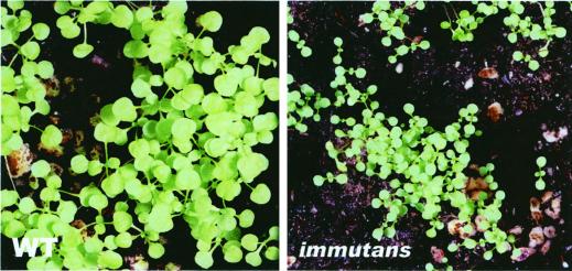
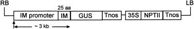
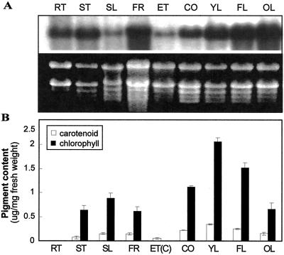
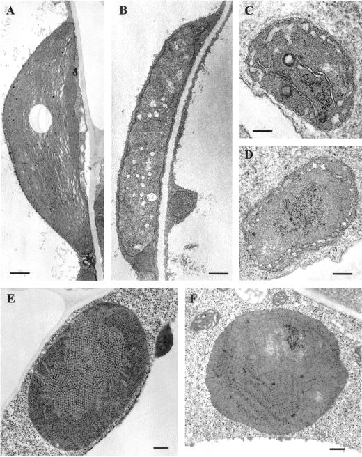
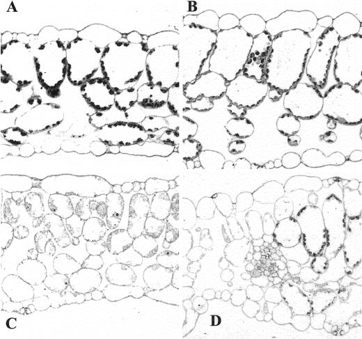
The immutans (im) variegation mutant of Arabidopsis has green and white leaf sectors due to the action of a nuclear recessive gene, IMMUTANS (IM). This gene encodes the IM protein, which is a chloroplast homolog of the mitochondrial alternative oxidase. Because the white sectors of im accumulate the noncolored carotenoid, phytoene, IM likely serves as a redox component in phytoene desaturation. In this paper, we show that IM has a global impact on plant growth and development and is required for the differentiation of multiple plastid types, including chloroplasts, amyloplasts, and etioplasts. IM promoter activity and IM mRNAs are also expressed ubiquitously in Arabidopsis. IM transcript levels correlate with carotenoid accumulation in some, but not all, tissues. This suggests that IM function is not limited to carotenogenesis. Leaf anatomy is radically altered in the green and white sectors of im: Mesophyll cell sizes are dramatically enlarged in the green sectors and palisade cells fail to expand in the white sectors. The green im sectors also have significantly higher than normal rates of O(2) evolution and elevated chlorophyll a/b ratios, typical of those found in "sun" leaves. We conclude that the changes in structure and photosynthetic function of the green leaf sectors are part of an adaptive mechanism that attempts to compensate for a lack of photosynthesis in the white leaf sectors, while maximizing the ability of the plant to avoid photodamage.


Figures

Figure 1
Figure 1
Growth of wild type and im. Plants were maintained under low-light conditions (15 μmol m−2 s−1) and photographed 8 weeks after germination. The wild type has an average of four true leaves and im an average of two true leaves. The seeds germinated at the same time. A magnification of 5× applies for both left and right panels.
Figure 2
Figure 2
Root growth in wild type and im. Root lengths were measured after 4 d of growth on Murashige and Skoog medium supplemented with 1% (w/v) Suc. The plants were maintained under normal light (100 μmol m−2 s−1), low light (15 μmol m−2 s−1), and in darkness. Each data point represents an individual plant.
Figure 3
Figure 3
pPZP/IMGUS, the IM promoter: GUS fusion construct. pPZP/IMGUS contains an approximately 3-kb upstream region of IM fused to the GUS (β-glucuronidase) gene and nos terminator. The selectable marker is an NPTII gene fused to 35S promoter/nos terminator elements. Twenty-five amino acids in the fusion protein are from the IM protein (Wu et al., 1999). RB, Right border; LB, left border.
Figure 4
Figure 4
Expression patterns of the IM promoter: GUS transgene during development. A, One-day-old light-grown seedlings. B, Seven-day-old light-grown seedling. C, Dark-field microscopy of a 4-d-old etiolated seedling (5× objective). D, Four-day-old etiolated seedling (35S promoter: GUS fusion; 5× objective). E, Cross-section of a shoot meristem of a 10-d-old light-grown seedling (25× objective). F and G, Cross-sections of first true leaves of 10-d-old light-grown seedlings (10× and 25× objectives, respectively). H, Six-week-old rosette. I, Bolt from a flowering plant. J, Cross-section of a hypocotyl of a 10-d-old light-grown seedling (25× objective). K, Root tip of a 10-d-old light-grown seedling. L, Dark-field microscopy of a flower (5× objective). M, Young seeds. AM, Apical meristem; AT, anther; COT, cotyledon; EP, epidermis; GC, guard cell; GT, ground tissues; HC, hypocotyl; MC, mesophyll cell; VT, vascular tissues; TR, trichome.
Figure 5
Figure 5
Expression analysis of IM mRNA and pigment levels in Arabidopsis. A, RNA gel-blot analyses were performed as described in “Materials and Methods.” The RNA gel is stained with ethidium bromide to show rRNA (loading control). The blot was probed with a radiolabeled IM cDNA (Wu et al., 1999). B, Total carotenoids and chlorophylls were extracted from Arabidopsis as described in “Materials and Methods.” Values are an average of three separate experiments ±sd. The samples in A and B are from 4- to 5-week-old plants grown under normal light conditions (100 μmol m−2 s−1), with the exception of the samples from dark-grown seedlings (ET). RT, Root; ST, stem; SL, green silique; FR, flowers (petals + green sepals); ET, 7-d-old etiolated seedling (cotyledon + hypocotyl); ET(C), cotyledons from 7-d-old etiolated seedlings; CO, 7-d-old cotyledon; YL, young leaf (5-mm length); FL, just fully expanded leaf (40-mm length); OL, senescing, late fully expanded leaf.
Figure 6
Figure 6
Plastid ultrastructure. Wild-type and im seedlings were grown on Murashige and Skoog plates for 7 d under normal light conditions (A, B, C, and D) or in darkness (E and F). A, Chloroplast from a wild-type cotyledon (bar = 500 nm). B, Chloroplast from an im cotyledon (bar = 500 nm). C, Amyloplast from a wild-type root (bar = 200 nm). D, Amyloplast from an im root (bar = 200 nm). E, Etioplast from a wild-type cotyledon (bar = 200 nm). F, Etioplast from an im cotyledon (bar = 200 nm).
Figure 7
Figure 7
Light microscopy of fully expanded leaves from wild-type and im plants grown under normal light conditions. A magnification of 25× applies to A through D. The white sectors stain less intensely than green sectors because their plastids are deficient in internal structures. A, Wild type. B, Green leaf sector of im. C, White leaf sector of im. D, Adjacent green and white sectors of im.
Figure 8
Figure 8
Photosynthetic oxygen evolution and chlorophyll a/b ratios in wild-type leaves and im green leaf sectors. Plants were grown under normal light (100 μmol m−2 s−1) or low light (15 μmol m−2 s−1). Oxygen evolution (A) and chlorophyll a/b ratios (B) were determined as described in “Materials and Methods.” Each graph represents an average ± sd of three different leaf samples for each illumination condition.

References

    1. Allen JF, Holmes NG. Electron transport and redox titration. In: Hipkins MF, Baker NR, editors. Photosynthesis Energy Transduction: A Practical Approach. Oxford: IRL Press; 1986. pp. 103–140.
    1. Bartley GE, Viitanen PV, Pecker I, Chamovitz D, Hirschberg J, Scolnik PA. Molecular cloning and expression in photosynthetic bacteria of a soybean cDNA coding for phytoene desaturase, an enzyme of the carotenoid biosynthesis pathway. Proc Natl Acad Sci USA. 1991;88:6532–6536. - PMC - PubMed
    1. Beyer P, Mayer M, Kleinig H. Molecular oxygen and the state of geometric isomerism of intermediates are essential in the carotene desaturation and cyclization reactions in daffodil chromoplasts. Eur J Biochem. 1989;184:141–150. - PubMed
    1. Bowman J. Arabidopsis: An Atlas of Morphology and Development. New York: Springer-Verlag; 1994.
    1. Carol P, Stevenson D, Bisanz C, Breitenbach J, Sandmann G, Mache R, Coupland G, Kuntz M. Mutations in the Arabidopsis gene IMMUTANS cause a variegated phenotype by inactivating a chloroplast terminal oxidase associated with phytoene desaturation. Plant Cell. 1999;11:57–68. - PMC - PubMed

Publication types

MeSH terms