. 2001 Sep 25;98(20):11312-7.
doi: 10.1073/pnas.191361798. Epub 2001 Sep 11.

SUMO-1 modification required for transformation by adenovirus type 5 early region 1B 55-kDa oncoprotein

Affiliations

SUMO-1 modification required for transformation by adenovirus type 5 early region 1B 55-kDa oncoprotein

C Endter et al. Proc Natl Acad Sci U S A. .

Abstract

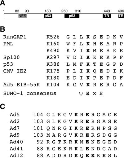
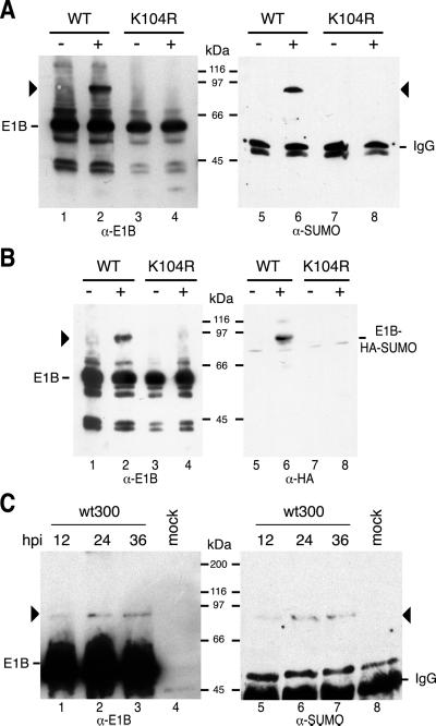
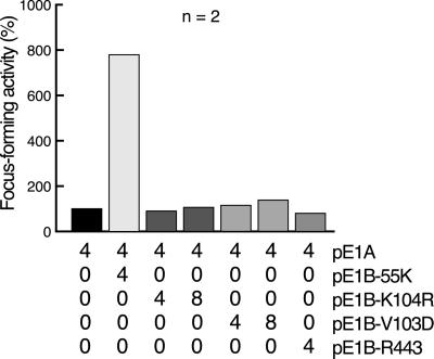
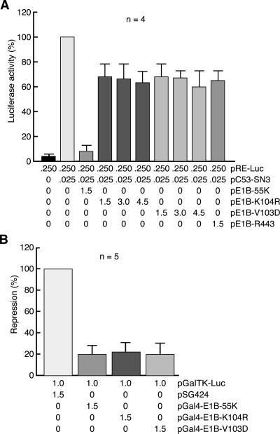
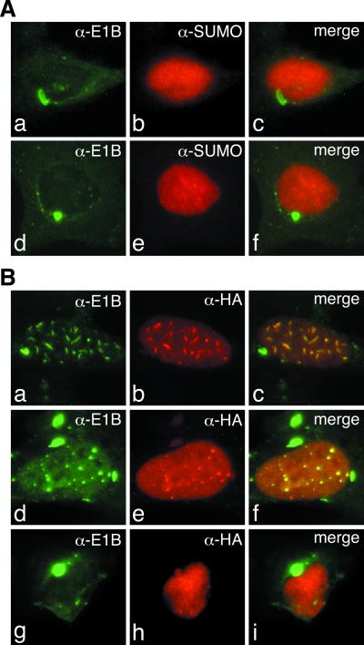
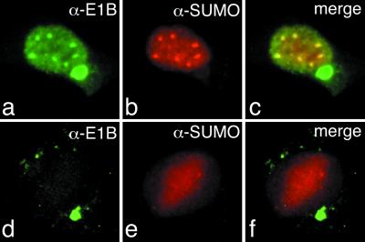
SUMO-1 is a small ubiquitin-related modifier protein that is covalently linked to many cellular and viral protein targets. Modification by SUMO-1 is proposed to play a role in protein targeting and/or stability. We show here that adenovirus type 5 early region 1B 55-kDa (E1B-55kDa) oncoprotein can be covalently modified by SUMO-1 in vivo through a major attachment site comprising a single lysine residue at amino acid position 104. The sequence surrounding this lysine matches the proposed PsiKxE consensus motif required for SUMO-1 conjugation. A single mutation (K104R) that abolishes SUMOylation of E1B-55kDa dramatically reduces the ability of the adenovirus type 5 protein to transform primary baby rat kidney cells in cooperation with E1A and to inhibit p53-mediated transactivation. Overexpression of SUMO-1 in adenovirus type 5 E1A/E1B-55kDa-transformed baby rat kidney cells causes the relocalization of E1B-55kDa from the cytoplasm to the nucleus, where it accumulates with SUMO-1 in dot- or track-like structures. Significantly, when SUMO-1 is ectopically expressed in transformed rat cells no effect on the cytoplasmic localization of the E1B-K104R mutant protein is observed. Our results demonstrate that SUMO-1 modification is required for transformation by adenovirus type 5 E1B-55kDa and provide further evidence for the idea that this posttranslational modification plays a role in protein targeting to specific subcellular sites.


Figures

Figure 1
Figure 1
(A) Diagram of Ad5 E1B-55kDa. Domains required for p53 binding (p53), transcriptional repression (TR), and transformation are indicated by black boxes. Numbers refer to amino acid residues, and 496 denotes the last amino acid. The location of the NES sequence is represented by a gray box. (B) Comparison of SUMO acceptor sites from several proteins with Ad5 E1B-55kDa. The consensus sequences of RanGAP1, PML, Sp100, p53, and cytomegalovirus (CMV) IE2 were derived from the sequences listed in ref. . Numbers indicate the position of the lysine residue (K) that serves as the SUMO attachment site; ψ stands for an aliphatic amino acid, and x represents any amino acid residue. (C) Alignment of potential SUMO acceptor sites in large E1B proteins from different human serotypes. Numbers indicate the position of the lysine residue (K) within the consensus motif.
Figure 2
Figure 2
Ad5 E1B-55kDa is covalently modified by SUMO-1 in vivo. (A) Subconfluent H1299 cells were transfected with pE1B-55K (WT) or pE1B-K104R (K104R) in the absence (−) or presence (+) of pSUMO-1. E1B-55kDa (E1B) and a slower-migrating form of the Ad protein indicated by black arrowheads were detected by the immunoblotting of total cell extracts with anti-E1B-55kDa mAb 2A6 (Left, α-E1B) or by immunoprecipitation of the same extracts with mAb 2A6 followed by immunoblotting with anti-SUMO-1 mAb 21C7 (Right, α-SUMO). (B) A similar high-molecular weight form of E1B-55kDa (Left, black arrowhead) was detected in total cell extracts prepared from H1299 cells transfected with pE1B-55K (WT) and pHA-SUMO-1 expressing an epitope-tagged SUMO-1 fusion protein. The high-molecular size band in lanes 2 and 6 corresponds to HA-SUMO-1-modified E1B-55kDa (E1B-HA-SUMO), as confirmed by Western blots with mAb 2A6 (Left, α-E1B) and anti-HA mouse mAb 12CA5 (Right, α-HA). (C) Covalent modification of E1B-55kDa by SUMO-1 in virus-infected cells. MCF7 cells were infected with wt300 virus, total cell extracts were prepared at the indicated time points in hours postinfection (hpi) and subjected to immunoprecipitation with mAb 2A6. Coprecipitated proteins were visualized by immunoblotting using mAbs 2A6 (Left, α-E1B) and 21C7 (Right, α-SUMO). E1B-55kDa (E1B) and the high-molecular weight form of E1B-55kDa (black arrowheads) are indicated. The positions of molecular mass markers are indicated in all panels.
Figure 3
Figure 3
Focus formation by Ad5 E1A, E1B-55kDa, E1B-K104R, E1B-V103D, and E1B-R443. Primary BRK cells were transfected with the indicated amounts of plasmids (micrograms of DNA per 3 × 10⁶ cells). Focus-forming activity is represented as a percentage of E1A alone. The average number of foci for pE1A was 3 in two independent experiments.
Figure 4
Figure 4
Effects of E1B-K104R, E1B-V103D, and E1B-R443 on p53 transcriptional activation. (A) Subconfluent H1299 cells were transfected with the indicated amounts of reporter and effector plasmids (micrograms of DNA). The mean and standard deviation are presented for four experiments, each performed in duplicate. (B) Effect of E1B-K104R and E1B-V103D on transcriptional repression. H1299 cells were transfected with the indicated amount of effector and reporter plasmids (micrograms of DNA). The mean and standard deviation are presented for five experiments, each performed in duplicate. pSG424 denotes the parental plasmid expressing the Gal4 DNA-binding domain under the control of the simian virus 40 early promoter (37).
Figure 5
Figure 5
(A) Subcellular localization of E1B-55kDa and SUMO-1 in transformed rat cells. AB120 and AB19 cells expressing wild-type E1B-55kDa (ac) or the K104R mutant protein (df), respectively, were double-labeled in situ with anti-E1B-55kDa rat mAb 9C10 or mouse mAb 21C7 specific for SUMO-1. These were detected with FITC- and Texas-red-conjugated secondary antibodies, respectively. Anti-E1B (green, a and d) and anti-SUMO-1 (red, b and e) staining patterns are shown. An overlay of these two patterns (merge) is shown in c and f. (B) Effect of ectopically expressed HA-SUMO-1 on the localization of wild-type E1B-55kDa and E1B-K104R. AB120 (af) and AB19 cells (gi) were transfected with 4 μg pHA-SUMO-1. After 36 h, cells were double-labeled with anti-E1B-55kDa mAb 2A6 (green, a, d, and g) and anti-HA rat mAb 3F10 (red, b, e, and h). The overlay of these patterns (merge) are shown in c, f, and i. (×7,600.)
Figure 6
Figure 6
Subcellular localization of E1B-55kDa and SUMO-1 in the presence of LMB. AB120 (ac) and AB19 cells (de) were grown for 3 h in medium containing 10 nM LMB before fixation. Then the cells were double-labeled with mAbs 9C10 (green, a and d) and 21C7 (red, b and e). The overlay of these images (merge) is shown in c and f. (×7,600.)

Similar articles

Cited by

References

    1. Shenk T. In: Virology. Fields B N, Knipe D M, Howley P M, editors. Vol. 2. New York: Lippincott-Raven; 1996. pp. 2111–2148.
    1. Barker D D, Berk A J. Virology. 1987;156:107–121. - PubMed
    1. Sarnow P, Ho Y S, Williams J, Levine A J. Cell. 1982;28:387–394. - PubMed
    1. Yew P R, Berk A J. Nature (London) 1992;357:82–85. - PubMed
    1. Teodoro J G, Branton P E. J Virol. 1997;71:3620–3627. - PMC - PubMed

Publication types

MeSH terms