. 2001 Sep 25;98(20):11714-9.
doi: 10.1073/pnas.191369798. Epub 2001 Sep 11.

Vitamin A deprivation results in reversible loss of hippocampal long-term synaptic plasticity

Affiliations

Vitamin A deprivation results in reversible loss of hippocampal long-term synaptic plasticity

D L Misner et al. Proc Natl Acad Sci U S A. .

Abstract

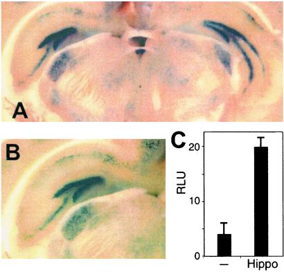
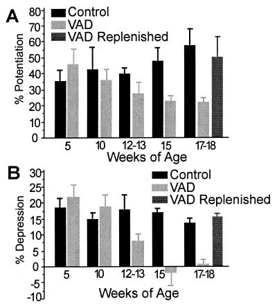
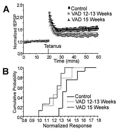
Despite its long history, the central effects of progressive depletion of vitamin A in adult mice has not been previously described. An examination of vitamin-deprived animals revealed a progressive and ultimately profound impairment of hippocampal CA1 long-term potentiation and a virtual abolishment of long-term depression. Importantly, these losses are fully reversible by dietary vitamin A replenishment in vivo or direct application of all trans-retinoic acid to acute hippocampal slices. We find retinoid responsive transgenes to be highly active in the hippocampus, and by using dissected explants, we show the hippocampus to be a site of robust synthesis of bioactive retinoids. In aggregate, these results demonstrate that vitamin A and its active derivatives function as essential competence factors for long-term synaptic plasticity within the adult brain, and suggest that key genes required for long-term potentiation and long-term depression are retinoid dependent. These data suggest a major mental consequence for the hundreds of millions of adults and children who are vitamin A deficient.


Figures

Figure 1
Figure 1
Retinoid detection in the adult hippocampus. (A and B) Two examples show X-Gal staining in the adult hippocampus of GAL4-RAR transgenic mice, giving rise to scattered, strongly positive cells in CA1, -2, and -3 of the hippocampus. More intense staining is seen in the dentate gyrus. Scattered positive cells are also present in the cortex and in undefined nuclei of the thalamus. (C) Adult hippocampal explants have been cocultured with JEG-3 cells transfected with GAL4-RAR expression vector and a GAL4 responsive luciferase reporter. The data demonstrate that hippocampus contains (low levels of) RAR-inducing activity, presumably retinoic acid.
Figure 2
Figure 2
Hippocampal CA1 LTP and LTD is impaired in VAD mice, but can be rescued by dietary vitamin A replenishment. (A) Summary of field potential LTP of control slices (black) at 5 weeks (37.2 ± 7.7%; n = 10), 10 weeks (44.9 ± 14.9%; n = 6), 12–13 weeks (41.6 ± 3.9%; n = 8), 15 weeks (50.0 ± 7.9%; n = 4), and 17–18 weeks (60.4 ± 11.6%; n = 10). Summary of field potential LTP of VAD slices (light gray) at 5 weeks (43.3 ± 9.0%; n = 8), 10 weeks (37.9 ± 7.5%; n = 12), 12–13 weeks (28.5 ± 7.5%; n = 9), 15 weeks (23.5 ± 4.4%; n = 8), and 17–18 weeks (23.4 ± 3.7%; n = 26). Summary of field potential LTP of VAD replenished (gray) slices at 17–18 weeks (52.7 ± 13.9%; n = 9). Data are expressed as the mean percent potentiation 25–35 min after tetanic stimulation ± SEM. (B) Summary of field potential LTD of control slices (black) at 5 weeks (19.5 ± 3.0%; n = 8), 10 weeks (15.7 ± 2.5%; n = 7), 12–13 weeks (18.8 ± 5.2%; n = 8), 15 weeks (17.7 ± 1.3%; n = 3), and 17–18 weeks (14.2 ± 1.7%; n = 8). Summary of field potential LTD of VAD slices (light gray) at 5 weeks (23.0 ± 3.9%; n = 9), 10 weeks (19.8 ± 4.3%; n = 11), 12–13 weeks (8.3 ± 2.4%; n = 7), 15 weeks (−1.9 ± 4.2%; n = 5), and 17–18 weeks (0.8 ± 1.6%; n = 11). Summary of field potential LTD of VAD replenished (gray) slices at 17–18 weeks (16.5 ± 1.5%; n = 6). Data are expressed as the mean percent depression 25–35 min after low frequency stimulation ± SEM.
Figure 3
Figure 3
LTP is impaired in VAD mice by 12–13 weeks of age. (A) Summary of field potential recordings from slices of control (squares; 1.499 ± 0.077; n = 12), VAD mice at 12–13 weeks of age (circles; 1.297 ± 0.071; n = 9), and VAD mice at 15 weeks of age (triangles; 1.229 ± 0.049; n = 8). The initial slope of fEPSPs is normalized to the baseline value preceding the induction of LTP. Data are expressed as mean ± SEM. Testing stimuli were given every 20 s. (B) Cumulative probability histogram of the magnitude of field potential LTP. Cumulative probability is shown as a function of mean response 25–35 min after tetanic stimulation.
Figure 4
Figure 4
LTD is severely impaired in VAD mice at 12–13 weeks and virtually abolished at 15 weeks. (A) Summary of field potential recordings from slices of control (squares; 0.83 ± 0.028; n = 11), VAD mice at 12–13 weeks of age (circles; 0.917 ± 0.024; n = 7), and VAD mice at 15 weeks of age (triangles; 1.019 ± 0.042; n = 5). The initial slope of fEPSPs is normalized to the baseline value preceding the induction of LTD. Data are expressed as mean ± SEM. Testing stimuli were given every 20 s. (B) Cumulative probability histogram of the magnitude of field potential LTD. Cumulative probability is shown as a function of mean response 25–35 min after low frequency stimulation.
Figure 5
Figure 5
Direct application of retinoic acid to VAD slices for ≥12 h rescues LTP. (A) Summary of field potential recordings from control slices (squares; 1.595 ± 0.12; n = 10), retinoic acid applied to VAD slices for <12 h (circles; 1.222 ± 0.033; n = 4), and retinoic acid applied to VAD slices for ≥12 h (triangles; 1.656 ± 0.179; n = 5). The initial slope of fEPSPs is normalized to the baseline value preceding the induction of LTP. Data are expressed as mean ± SEM. Testing stimuli were given every 20 s. (B) Cumulative probability histogram of the magnitude of field potential LTP. Cumulative probability is shown as a function of mean response 25–35 min after tetanic stimulation.
Figure 6
Figure 6
Direct application of retinoic acid to VAD slices for ≥12 h rescues LTD. (A) Summary of field potential recordings from control slices (squares; 0.854 ± 0.019n = 8), retinoic acid applied to VAD slices for <12 h (circles; 0.96 ± 0.035; n = 3), and retinoic acid applied to VAD slices for ≥12 h (triangles; 0.826 ± 0.068; n = 5). The initial slope of fEPSPs is normalized to the baseline value preceding the induction of LTD. Data are expressed as mean ± SEM. Testing stimuli were given every 20 s. (B) Cumulative probability histogram of the magnitude of field potential LTD. Cumulative probability is shown as a function of mean response 25–35 min after low frequency stimulation.
Figure 7
Figure 7
Overall hippocampal structure is maintained in VAD mice. Coronal slices of hippocampus in control (A) and VAD (B) mice show indistinguishable morphology, indicating that vitamin A deficiency does not result in obvious neurodegeneracy of hippocampus.

References

    1. Mangelsdorf D J, Umesono K, Evans R M. In: The Retinoids: Biology, Chemistry, and Medicine. 2nd Ed. Sporn M B, Roberts A B, Goodman D S, editors. New York: Raven; 1994. pp. 319–349.
    1. Krezel W, Kastner P, Chambon P. Neuroscience. 1999;89:1291–1300. - PubMed
    1. Underwood B A, Arthur P. FASEB J. 1996;10:1040–1048. - PubMed
    1. Chambon P. Semin Cell Biol. 1994;5:115–125. - PubMed
    1. Kastner P, Mark M, Ghyselinck N, Krezel W, Dupe V, Grondona J M, Chambon P. Development (Cambridge, UK) 1997;124:313–326. - PubMed

Publication types

MeSH terms