. 2001 Sep 11;98(19):10602-7.
doi: 10.1073/pnas.191268798.

Structural features of an influenza virus promoter and their implications for viral RNA synthesis

Affiliations

Structural features of an influenza virus promoter and their implications for viral RNA synthesis

S H Bae et al. Proc Natl Acad Sci U S A. .

Abstract

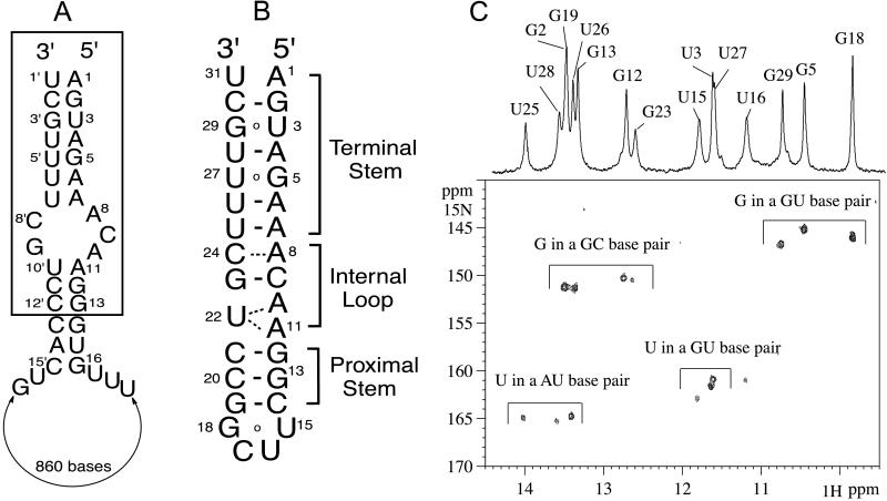
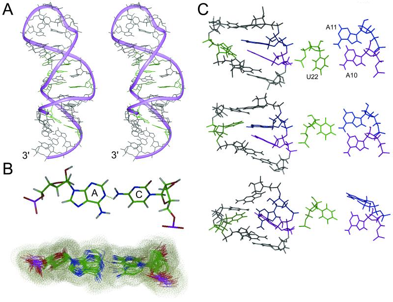
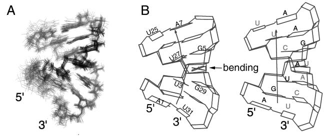
The influenza A virus, a severe pandemic pathogen, has a segmented RNA genome consisting of eight single-stranded RNA molecules. The 5' and 3' ends of each RNA segment recognized by the influenza A virus RNA-dependent RNA polymerase direct both transcription and replication of the virus's RNA genome. Promoter binding by the viral RNA polymerase and formation of an active open complex are prerequisites for viral replication and proliferation. Here we describe the solution structure of this promoter as solved by multidimensional, heteronuclear magnetic resonance spectroscopy. Our studies show that the viral promoter has a significant dynamic nature and reveal an unusual displacement of an adenosine that forms a novel (A-A) x U motif and a C-A mismatch stacked in a helix. The characterized structural features of the promoter imply that the specificity of polymerase binding results from an internal RNA loop. In addition, an unexpected bending (46 +/- 10 degrees ) near the initiation site suggests the existence of a promoter recognition mechanism similar to that of DNA-dependent RNA polymerase and a possible regulatory function for the terminal structure during open complex formation.


Figures

Figure 1
Figure 1
(A) Terminal sequence of the influenza A virus genomic RNA. The conserved 13 nt at the 5′ end and 12 nt at the 3′ ends are boxed. The numbering from the 3′ end is followed by a prime notation (′). The sequence shown is that of viral RNA (vRNA) segment 8 of influenza A/PR/8/34. (B) Secondary structure of the RNA construct used in this study. Watson–Crick and non-Watson–Crick base pairs are distinguished by bars and circles, respectively. Dashed lines indicate the calculation-driven base pairs. (C) Imino region of 1H-15N HSQC spectrum at 278 K.
Figure 2
Figure 2
(A) The overall structure of the influenza A virus promoter. A4⋅U28 in the terminal stem, A8⋅C24 mismatch, and the (A-A)⋅U motif in the internal loop are shown in green. (B) A potential hydrogen bond formed between A8 and C24 (up) and superposition of 32 structures of the A⋅C mismatch (down). (C) Three groups of low-energy structures of the (A-A)⋅U motif are shown, viewed from the major groove (Left) and from the top (Right). A10 is shown in purple, A11 in blue, and U22 in green.
Figure 3
Figure 3
(A) The pyrimidine H5 to H6 region of an 80-ms NOESY spectrum (green-blue) superimposed on the DQF-COSY spectrum (orange-red). Each H5-H6 cross peak of pyrimidines was assigned on the NOESY spectrum. (B) The selected NOE cross peaks of H5 and H6 of pyrimidine residues obtained from 400 MHz and 800 MHz NOESY spectra at 294 K. (C) The H2 to H1′ region of a 150-ms NOESY spectrum recorded at 294 K. The H2 resonances of A10 and A11 are indicated by a horizontal dashed lines, and the H1′ resonances of A11, G12, and G23 are indicated by a vertical dashed line. The red circled cross peak indicates the unusual NOE between A10H2 and G23H1′.
Figure 4
Figure 4
(A) The 32 converged structures, superimposed on the terminal stem. (B) Schematic representation of the helical geometry of the terminal stem (Left) and of a normal A-form helix (Right) with a complete Watson–Crick base pair (5′-AGUAGAA/3′-UCAUCUU). The one-headed arrow indicates the helical axis. This figure was generated with CURVES 5.2.

References

    1. Lamb R A, Choppin P W. Annu Rev Biochem. 1983;52:467–506. - PubMed
    1. Luytjes W, Krystal M, Enami M, Pavin J D, Palese P. Cell. 1989;59:1107–1113. - PubMed
    1. Fodor E, Pritlove D C, Brownlee G G. J Virol. 1994;68:4092–4096. - PMC - PubMed
    1. Fodor E, Pritlove D C, Brownlee G G. J Virol. 1995;69:4012–4019. - PMC - PubMed
    1. Cianci C, Tiley L, Krystal M. J Virol. 1995;69:3995–3999. - PMC - PubMed

Publication types

MeSH terms

Associated data

LinkOut - more resources