. 2001 Sep 11;98(19):10728-32.
doi: 10.1073/pnas.181336098.

Rhesus monkey placental transgene expression after lentiviral gene transfer into preimplantation embryos

Affiliations

Rhesus monkey placental transgene expression after lentiviral gene transfer into preimplantation embryos

M J Wolfgang et al. Proc Natl Acad Sci U S A. .

Abstract

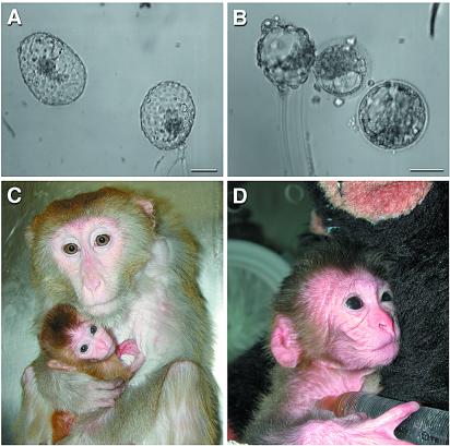
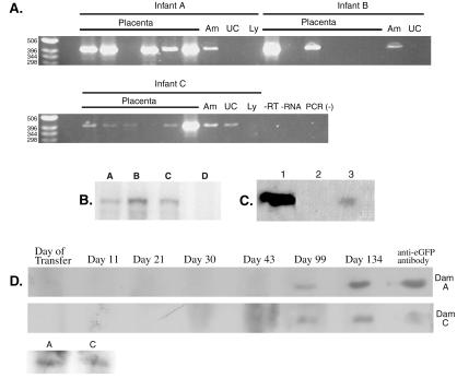
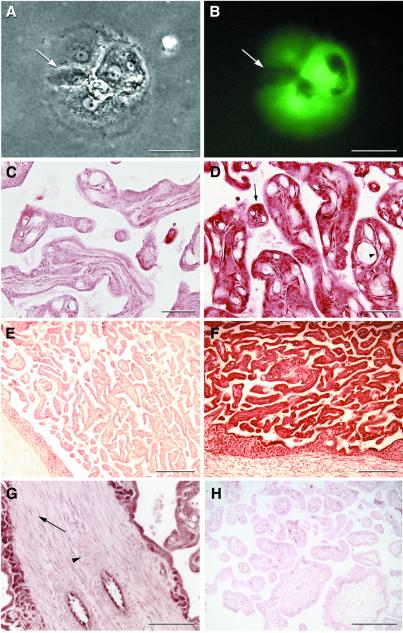
Transgenic mice have provided invaluable information about gene function and regulation. However, because of marked differences between rodents and primates, some areas of human biology such as early embryonic development, aging, and maternal-fetal interactions would be best studied in a nonhuman primate model. Here, we report that gene transfer into rhesus monkey (Macaca mulatta) preimplantation embryos gives rise to transgenic placentas that express a reporter transgene (eGFP). Blastocysts resulting from culture of in vitro fertilized ova were transduced with a self-inactivating lentiviral vector and transferred into recipient females. One twin and one singleton pregnancy were produced from a single stimulation cycle, and one live rhesus monkey was born from each pregnancy. Placentas from all conceptuses showed expression of the transgene as detected by reverse transcription-PCR, ribonuclease protection assay, direct epifluorescence, immunohistochemistry, and Western blot analysis. Integration in somatic tissues of the offspring was not detected. A maternal immune response to the xenogeneic placental antigen was shown by the presence of anti-GFP antibodies in peripheral blood of the recipient females by day 99 of gestation (term = 165 days). These results demonstrate that transgene expression during gestation is compatible with successful pregnancy in nonhuman primates and provides an approach that could be broadly applicable to the development of novel models for primate biomedical research.


Figures

Figure 1
Figure 1
SIN lentiviral vector-transduced rhesus monkey blastocysts and infants. In vitro produced blastocysts that contributed to the twin and singleton infants, respectively (A and B). Shown are the surviving twin (infant A) with dam (C) and male singleton (infant C) in a perinatal incubator holding a 10% dextrose syringe with nipple (D). He subsequently was returned to his dam as well. Embryos were exposed to greater than 1 sec of continuous epifluorescent excitation at 488 nm but were not yet expressing robust amounts of eGFP. (Bars = 100 μm.)
Figure 2
Figure 2
Transgene expression and the subsequent maternal humoral response. (A) RT-PCR in the three animals; results are shown for six placental biopsies along with amnion (Am), umbilical cord (UC), and cord lymphocytes (Ly). (B) Ribonuclease protection assay for eGFP mRNA. Lanes: A–C, placental RNA from animals A, B, and C; D, nontransgenic placenta. (C) Western blot for GFP. Lanes: 1, 100 ng recombinant eGFP; 2, control placenta; 3, 250 μg of total protein from placenta A. (D) Maternal serum samples from dam A and C as well as fetal cord blood (A and C, bottom blot) were assayed for antibodies directed against placental eGFP as detected by Western blot analysis. A positive control is shown that uses a mouse monoclonal anti-GFP antibody.
Figure 3
Figure 3
Transgene protein expression in placenta. Phase-contrast (A) and epifluorescent images (B) of cultured trophoblasts from placenta A are shown. Arrow indicates a nonexpressing trophoblast that has fused recently with a transgenic syncytium. (C and E) Bright-field negative controls for D and F. Anti-GFP immunohistochemistry of placenta A (D) frozen sections reveals widespread eGFP expression. Arrow indicates an expressing trophoblast, and arrowhead shows an expressing fetal villous endothelial cell. (F) Low-power view of placenta C showing eGFP expression throughout the trophoblastic shell. (G) Immunohistochemical localization of eGFP in placenta A showing the absence of expression in placental stroma and vascular smooth muscle. Arrow shows nonexpressing stromal cells, and arrowhead shows nonexpressing vascular smooth muscle cells. (H) Photomicrograph of a nontransgenic control placenta probed with the same anti-GFP antibody. [Bars = 50 μm (A and B), 40 μm (C and D), 200 μm (E, F, and H), and 100 μm (G).]

References

    1. Soriano P, Cone R D, Mulligan R C, Jaenisch R. Science. 1986;234:1409–1413. - PubMed
    1. Haskell R E, Bowen R A. Mol Reprod Dev. 1995;40:386–390. - PubMed
    1. Chan A W, Homan E J, Ballou L U, Burns J C, Bremel R D. Proc Natl Acad Sci USA. 1998;95:14028–14033. - PMC - PubMed
    1. Chan A W S, Chong K Y, Martinovich C, Simerly C, Schatten G. Science. 2001;291:309–312. - PubMed
    1. Hawley R G, Covarrubias L, Hawley T, Mintz B. Proc Natl Acad Sci USA. 1987;84:2406–2410. - PMC - PubMed

Publication types

Substances

LinkOut - more resources