

Dissolution of calcium pyrophosphate crystals by polyphosphates: an in vitro and ex vivo study
- PMID: 11557654
- PMCID: PMC1753396
- DOI: 10.1136/ard.60.10.962
Dissolution of calcium pyrophosphate crystals by polyphosphates: an in vitro and ex vivo study
Abstract
Objective: To determine the dissolving ability (DA) of linear pentasodium tripolyphosphate (PSTP), cyclic trisodium metaphosphate (TSMP), polymeric sodium metaphosphate (SMP) on synthetic crystals of calcium pyrophosphate dihydrate (CPPD) and on crystalline aggregates of menisci from patients with chondrocalcinosis (CC).
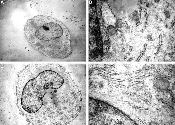
Methods: Synthetic CPPD crystals were mixed with phosphate buffered saline (PBS), which contained the different polyphosphates, for one hour at 37 degrees C. The calcified menisci were obtained from the knees of four female patients with CPPD disease who underwent total arthroscopic meniscectomy for degenerative meniscal lesions. Meniscal cryosections and fragments were incubated in SMP (15 mg/ml PBS) at 37 degrees C for one hour and 24 hours, respectively. Histological evaluation on meniscal samples after polyphosphate incubation was carried out by ordinary transmitted light microscopy and polarised light microscopy. The dissolution of CPPD crystals by polyphosphates was assessed by atomic absorption spectroscopy, which determined the amount of calcium liberated from synthetic crystals and meniscal fragments. Cytotoxicity of SMP was evaluated by tetrazolium salt assay and by an ultrastructural study on cultured chondrocytes.
Results: SMP and PSTP showed higher DA on CPPD crystals than TSMP. Analysis of the DA values at increasing concentrations of SMP showed that a concentration of 15 mg/ml completely dissolved 2.0 mg CPPD crystals. The solution of meniscal CPPD crystals showed a significant increase of calcium concentration after three hours and 24 hours of SMP incubation (p=0.0001; Kruskal-Wallis analysis of variance) compared with fragments incubated in PBS control solution. Macroscopic and microscopic evaluation of meniscal specimens showed a notable reduction of CPPD deposits. A 50% inhibitory dose on cultured chondrocytes was reached at the maximum concentration of SMP used in this work (15 mg/ml); ultrastructural analysis did not show morphological alterations in the treated cells.
Conclusion: The results of this study indicate that linear polyphosphates are effective in dissolving both synthetic and ex vivo CPPD crystal aggregates. This suggests a potential therapeutic use for these molecules in the treatment of symptomatic CC.
Figures


References
Publication types
MeSH terms
Substances
LinkOut - more resources
Full Text Sources
Other Literature Sources
