

Tibial and femoral cartilage changes in knee osteoarthritis
- PMID: 11557657
- PMCID: PMC1753397
- DOI: 10.1136/ard.60.10.977
Tibial and femoral cartilage changes in knee osteoarthritis
Abstract
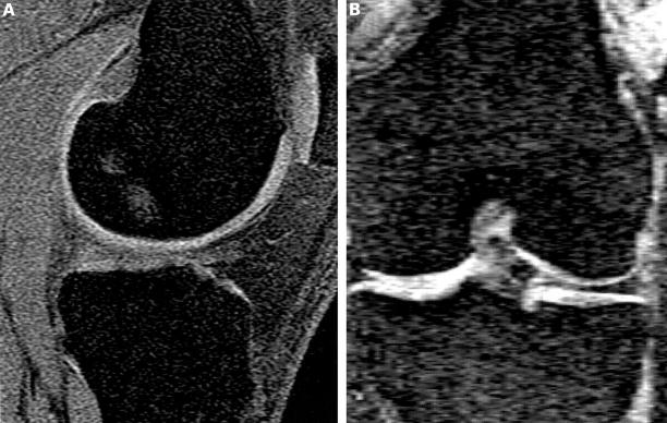
Background: Despite the increasing interest in using knee cartilage volume as an outcome measure in studies of osteoarthritis (OA), it is unclear what components of knee cartilage will be most useful as markers of structure in the tibiofemoral (TF) joint.
Objective: To compare the changes that occur in femoral and tibial cartilage volume in normal and osteoarthritic knees and how they relate to radiological grade.
Methods: 82 subjects (44 female, 38 male, age range 35-69 years) with a spectrum of radiological knee OA were examined. Each subject had femoral and tibial cartilage volume in the medial and lateral TF joint determined from T(1) weighted fat saturated magnetic resonance images of the knee. Radiological grade of OA was determined from standing knee radiographs.
Results: There was strong correlation between femoral and tibial cartilage volume measured in both the medial (R=0.75, p<0.001) and lateral TF joint (R=0.77, p<0.001). Similar correlations persisted when those with normal and those with OA joints were examined separately at both the medial and lateral TF joint. For each increase in radiological grade of joint space narrowing (0-3), there was a mean (SD) reduction in tibial cartilage volume of 1.00 (0.32) ml in the medial compartment and 0.53 (0.25) ml in the lateral compartment, after adjusting for differences in bone size. Similar changes were seen in the femoral cartilage.
Conclusions: The amounts of tibial and femoral cartilage are strongly related. It may be that for TF joint disease, measuring tibial cartilage alone may be adequate, given that measurements of the total femoral cartilage are less reproducible and the difficulties inherent in identifying the most appropriate component of femoral cartilage to measure.
Figures
Publication types
MeSH terms
LinkOut - more resources
Full Text Sources
Other Literature Sources
Medical
Miscellaneous
