. 2001 Sep 15;29(18):3882-91.
doi: 10.1093/nar/29.18.3882.

Peptide-mediated RNA delivery: a novel approach for enhanced transfection of primary and post-mitotic cells

Affiliations

Peptide-mediated RNA delivery: a novel approach for enhanced transfection of primary and post-mitotic cells

T Bettinger et al. Nucleic Acids Res. .

Abstract

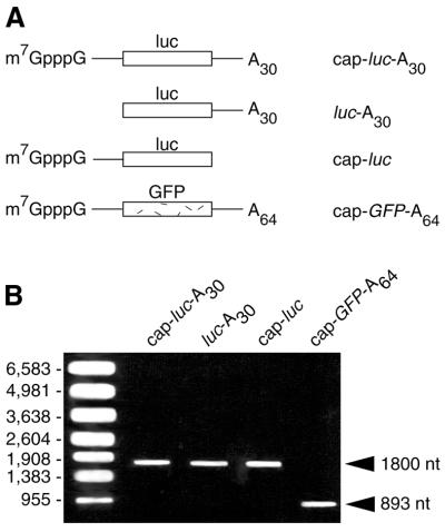
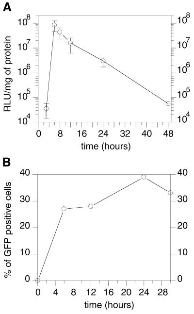
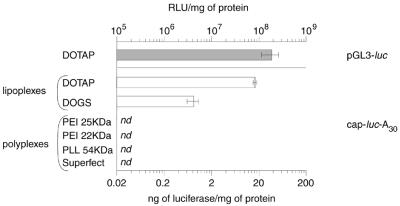
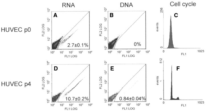
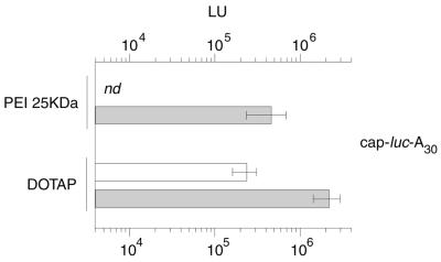
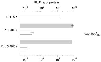
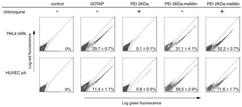
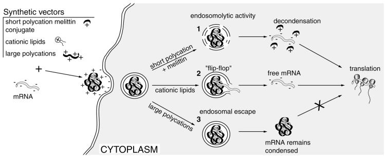
Synthetic vectors were evaluated for their ability to mediate efficient mRNA transfection. Initial results indicated that lipoplexes, but not polyplexes based on polyethylenimine (PEI, 25 and 22 kDa), poly(L-lysine) (PLL, 54 kDa) or dendrimers, mediated efficient translation of mRNA in B16-F10 cells. Significant mRNA transfection was achieved by lipoplex delivery in quiescent (passage 0) human umbilical vein endothelial cells (HUVEC), and by passage 4, 10.7% of HUVEC were transfected compared to 0.84% with DNA. Lack of expression with PEI 25 kDa/mRNA or PLL 54 kDa/mRNA in a cell-free translation assay and following cytoplasmic injection into Rat1 cells indicated that these polyplexes were too stable to release mRNA. In contrast, polyplexes formed using smaller PEI 2 kDa and PLL 3.4 kDa gave 5-fold greater expression in B16-F10 cells compared to DOTAP, but were dependent on chloroquine for transfection activity. Endosomolytic activity was incorporated by conjugating PEI 2 kDa to melittin and resulting PEI 2 kDa-melittin/mRNA polyplexes mediated high transfection levels in HeLa cells (31.1 +/- 4.1%) and HUVEC (58.5 +/- 2.9%) in the absence of chloroquine, that was potentiated to 52.2 +/- 2.7 and 71.6 +/- 1.7%, respectively, in the presence of chloroquine. These results demonstrate that mRNA polyplexes based on peptide-modified low molecular weight polycations can possess versatile properties including endosomolysis that should enable efficient non-viral mRNA transfection of quiescent and post-mitotic cells.


Figures

Figure 1
Figure 1
(A) Schematic representation of mRNAs used in this study. The in vitro transcripts were capped (m7GpppG) or uncapped, with or without a poly(A) tail, and encode for either luciferase or GFP. (B) RNA transcribed in vitro was denaturated by heating at 65°C for 15 min and analysed on a ethidium bromide-stained 0.8% agarose gel (2 µg of purified RNA per lane) and visualised by UV translumination. No differences in migration of mRNA-luc transcripts with or without poly(A) tail were observed under these conditions.
Figure 2
Figure 2
Time course of expression of mRNA transfection in B16-F10 cells. Complexes were prepared by mixing 1 µg of cap-luc-A30 with 5.4 nmol of DOTAP in HEPES buffer pH 7.4 as described in Materials and Methods. After 4 h post-transfection, 500 µl of fresh media containing 10% FCS were added to each well. Luciferase (A) or GFP (B) expression were assayed over 48 h and 30 h post-transfection, respectively.
Figure 3
Figure 3
Lipoplexes or polyplexes/mRNA-mediated transfection of B16-F10 cells. cap-luc-A30 (1 µg) condensed by cationic lipids (DOTAP N/P = 1.8, DOGS six charge equivalent) or cationic polymers (PEI 25 kDa N/P = 10, PEI 22 kDa N/P = 5, PLL 54 kDa N/P = 2 and Superfect N/P = 8.5) were delivered to B16-F10 cells and luciferase gene expression evaluated 6 h post-transfection. The efficiency of transfection was compared to the pGL3luc/DOTAP lipoplexes (luciferase expression measured 24 h post-transfection). Each experiment was performed in triplicate. nd, not detected.
Figure 4
Figure 4
Relative efficiency of DNA and mRNA-mediated transfection in quiescent and slow dividing HUVEC. DOTAP/cap-GFP-A64 and DOTAP/pEGFP complexes were delivered, respectively, to p0 (A and B) and p4 (D and E) HUVEC. Cells were plated in 48-well plates coated with collagen, and GFP expression measured after 24 h by flow cytometry analysis as described in Materials and Methods. The cell cycle of HUVEC at passage 0 and 4 were determined by the use of SYTOX Green.
Figure 5
Figure 5
In vitro translation of mRNA using rabbit reticulocyte lysate. cap-luc-A30 (1 µg) was incubated free or condensed by PEI 25 kDa at N/P = 10, and DOTAP at N/P = 1.8 with nuclease-treated rabbit reticulocyte lysate and luciferase activity was measured after 90 min. Luciferase expression for free cap-luc-A30 is 1.4 × 10⁷ LU. To determine whether the stability of lipoplexes and polyplexes influences the translatibility of cap-luc-A30. 100 µg/ml of polyaspartic acid (pAsp) was added to the rabbit reticulocyte lysate prior to PEI 25 kDa and DOTAP/RNA complexes (white bars without pAsp, shaded bars with 100 µg/ml pAsp). nd, not detected.
Figure 6
Figure 6
Transfection efficiency in B16-F10 cells of short polycation/mRNA complexes. B16-F10 cells were transfected with 1 µg of cap-luc-A30 condensed either by DOTAP at N/P = 1.8, or PEI 2 kDa at N/P = 5 or PLL 3.4 kDa at N/P = 7.5 in DMEM without serum in the absence (white bars) or presence (shaded bars) of 100 µM chloroquine. The cells were incubated with the complexes for 4 h and the medium discarded and replaced with fresh medium containing 10% FCS. Luciferase activity was measured after 6 h. Results are shown as a mean and standard deviation from triplicate samples.
Figure 7
Figure 7
Transfection efficiency of PEI 2 kDa–melittin conjugate in HeLa cells and HUVEC. HeLa cells and passage 4 HUVEC were transfected with 1 µg of cap-GFP-A64 condensed either by DOTAP at N/P = 1.8, or PEI 2 kDa at N/P = 5 or PEI 2 kDa–melittin at N/P = 5 in DMEM with serum. In some cases, cells were treated with 100 µM chloroquine when transfected with PEI 2 kDa and with PEI 2 kDa–melittin. GFP expression was measured after 24 h by flow cytometry analysis as described in Materials and Methods.
Figure 8
Figure 8
Proposed intracellular fate of delivered mRNA. The availability of mRNA to undergo translation in the cytoplasm is dependent on the type of synthetic vector used. (1) When delivered with short polycation containing the endosomolytic peptide melittin (PEI 2 kDa–melittin), complexes escape from the endosome and mRNA is efficiently released for translation to occur. In the case of unmodified short polycations (PEI 2 kDa, PLL 3.4 kDa), endosomal escape can be triggered by the buffering agent chloroquine. (2) When delivered with cationic lipids (DOTAP, DOGS), mRNA is released into the cytoplasm following a ‘flip-flop’ process between the cationic lipids and the anionic component of the endosomal membrane. (3) When larger polycations are used, polyplexes escape from the endosome either by release with chloroquine (PLL 54 kDa) or by the ‘proton sponge’ effect (PEI 25 kDa) (34). However, in the cytoplasm the polyplexes do not adequately release mRNA in a form suitable for translation to occur.

References

    1. Malone R.W., Felgner,P.L. and Verma,I.M. (1989) Cationic liposome-mediated RNA transfection. Proc. Natl Acad. Sci. USA, 86, 6077–6081. - PMC - PubMed
    1. Pollard H., Remy,J.S., Loussouarn,G., Demolombe,S., Behr,J.P. and Escande,D. (1998) Polyethylenimine but not cationic lipids promotes transgene delivery to the nucleus in mammalian cells. J. Biol. Chem., 273, 7507–7511. - PubMed
    1. Brunner S., Sauer,T., Carotta,S., Cotten,M., Saltik,M. and Wagner,E. (2000) Cell cycle dependence of gene transfer by lipoplex, polyplex and recombinant adenovirus. Gene Ther., 7, 401–407. - PubMed
    1. Nair S.K., Boczkowski,D., Morse,M., Cumming,R.I., Lyerly,H.K. and Gilboa,E. (1998) Induction of primary carcinoembryonic antigen (CEA)-specific cytotoxic T lymphocytes in vitro using human dendritic cells transfected with RNA. Nat. Biotechnol., 16, 364–369. - PubMed
    1. Strobel I., Berchtold,S., Gotze,A., Schulze,U., Schuler,G. and Steinkasserer,A. (2000) Human dendritic cells transfected with either RNA or DNA encoding influenza matrix protein M1 differ in their ability to stimulate cytotoxic T lymphocytes. Gene Ther., 7, 2028–2035. - PubMed

Publication types

MeSH terms