

Case Reports
Decreased diffusion in central pontine myelinolysis
Affiliations
- PMID: 11559493
- PMCID: PMC7974564
Item in Clipboard
Case Reports
Decreased diffusion in central pontine myelinolysis
AJNR Am J Neuroradiol.
2001 Sep.
Abstract
Two patients with central pontine myelinolysis (CPM) were studied with diffusion-weighted MR imaging 1 week after onset of tetraplegia. In both patients, affected white matter showed hyperintensity on diffusion-weighted images associated with a decrease in apparent diffusion coefficient (ADC) values. In one patient studied serially, ADC values normalized by 3 weeks after tetraplegia. Early in the clinical course, diagnosis of CPM can sometimes be difficult. Hyperintensity on diffusion-weighted images may therefore have diagnostic utility. Decreased lesional ADC values support the notion that CPM is a consequence of relative intracellular hypotonicity.
Figures
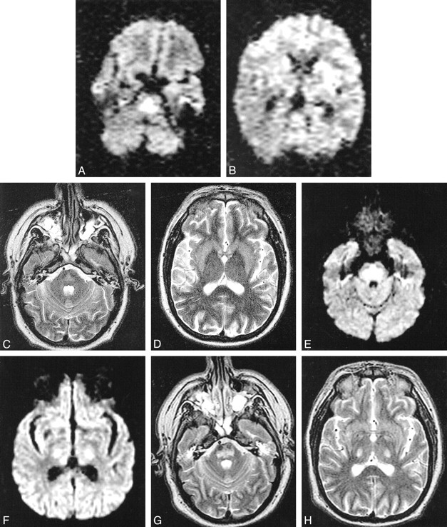
Case 1. MR images obtained using a 1.5-T magnet. Images for study 1 (A–D) were taken in an oblique axial plane 6 days after development of tetraplegia. Images for study 2 (E–H) were taken in an axial plane 21 days after development of tetraplegia. A, DW image shows increased signal intensity in pons due to restricted water diffusion. B, DW image is normal in the internal capsules bilaterally. C and D, In the pons, there is little hyperintensity on the T2-weighted images and low ADC values, with mean ADC in basis pontis being 0.39 ± 0.14 × 10⁻³ mm2/s (mean ± SD) compared with 0.90 ± 0.15 × 10⁻³ mm2/s in unaffected white matter. E, DW image shows increased signal intensity still present in the pons. F, The DW image shows increased signal intensity present in the internal capsules bilaterally. G and H, The DW abnormality is accounted for by shine-through from the hyperintense T2-weighted images. Mean ADC in basis pontis (1.09 ± 0.10 × 10⁻³ mm2/s) is normal compared with that of unaffected white matter (1.05 ± 0.09 × 10⁻³ mm2/s).
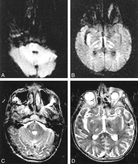
Case 2. Images obtained in the axial plane 7 days after development of tetraplegia. DW (A and B) and T2-weighted (C and D) images show increased signal intensity within the pons and the thalami bilaterally, consistent with CPM with extrapontine involvement. ADC is decreased in affected pontine (0.62 ± 0.11 × 10⁻³ mm2/s) and thalamic (0.43 ± 0.09 × 10⁻³ mm2/s) regions compared with that of unaffected white matter (0.82 ± 0.12 × 10⁻³ mm2/s). The patient died before repeat images could be obtained
References
-
- Adams RA, Victor M, Mancall EL. Central pontine myelinolysis: a hitherto undescribed disease occurring in alcoholics and malnourished patients. Arch Neurol Psychiat 1959;81:154-172 - PubMed
-
- McKee AC, Winkelman MD, Banker BQ. Central pontine myelinolysis in severely burned patients: relationship to serum hyperosmolality [published erratum appears in Neurology 1988;38:1662]. Neurology 1988;38:1211-1217 - PubMed
-
- Karp BI, Laureno R. Pontine and extrapontine myelinolysis: a neurologic disorder following rapid correction of hyponatremia. Medicine (Baltimore) 1993;72:359-373 - PubMed
-
- Schaefer PW, Grant PE, Gonzalez RG. Diffusion-weighted MR imaging of the brain. Radiology 2000;217:331-345 - PubMed
Publication types
MeSH terms
LinkOut - more resources
Full Text Sources
Medical