Case Reports
. 2001 Sep;22(8):1480-2.

Wernicke's encephalopathy: atypical manifestation at MR imaging

Affiliations
Case Reports

Wernicke's encephalopathy: atypical manifestation at MR imaging

S J Bae et al. AJNR Am J Neuroradiol. 2001 Sep.

Abstract

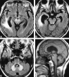
We report a case of atypical manifestation of hyperintense lesions in a 64-year-old female patient with Wernicke's encephalopathy. Fluid-attenuated inversion recovery and T2-weighted images demonstrated symmetrical distribution of hyperintense lesions in cerebellar dentate nuclei, tegmentum of the lower pons, red nuclei, and tectum of the midbrain, and T1-weighted sagittal images showed atrophy of the mamillary bodies. The hyperintense lesions were completely resolved on follow-up MR images.


Figures

<sc>fig</sc> 1.
fig 1.
March 22. A, Contrast-enhanced T1-weighted axial image shows abscess cavity (arrows) in the right peritrigonal area. B, T1-weighted sagittal image shows normal-appearing mamillary body (arrow).
<sc>fig</sc> 2.
fig 2.
May 29. A–C, FLAIR axial images. A, Red nuclei of the midbrain are hyperintense compared with the brain parenchyma (solid arrows). White matter hyperintensity (open arrow) adjacent to the dilated right lateral ventricle is probably a change due to the abscess cavity. B, Tectum of the midbrain also shows symmetric hyperintensities (arrows). C, At the lower pons level, symmetrical hyperintense lesions are also detected in facial nuclei (solid arrows), vestibular nuclei (open arrows), and abducens nuclei (arrowheads) of the pontine tegmentum and cerebellar dentate nuclei (double arrows). D, T1-weighted sagittal image shows atrophy of mamillary body (arrow).
<sc>fig</sc> 3.
fig 3.
July 10. A–C, FLAIR axial images show complete resolution of the previously detected unusual hyperintense lesions. Residual white matter hyperintensity is noted along the dilated right lateral ventricle.

References

    1. Doraiswamy PM, Massey EW, Enright K, Palese VJ, Lamonica D, Boyko O. Wernicke-Korsakoff syndrome caused by psychogenic food refusal: MR findings. AJNR Am J Neuroradiol 1994;15:594-596 - PMC - PubMed
    1. Opdenakker G, Gelin G, De Surgeloose D, Palmers Y. Wernicke encephalopathy: MR findings in two patients. Eur Radiol 1999;9:1620-1624 - PubMed
    1. Gallucci M, Bozzao A, Splendiani A, Masciocchi C, Passariello R. Wernicke encephalopathy: MR findings in five patients. AJNR Am J Neuroradiol 1990;11:887-892 - PMC - PubMed
    1. Yoon HK, Chang KH, Lee G, et al. MR findings of Wernicke encephalopathy. J Korean Radiol Soc 1991;27:485-491
    1. Suzuki S, Ichijo M, Fujii H, Matsuoka Y, Ogawa Y. Acute Wernicke's encephalopathy: comparison of magnetic resonance images and autopsy findings. Intern Med 1996;35:831-834 - PubMed

Publication types