. 2001 Sep;22(8):1510-6.

CT and MR imaging features of pyogenic ventriculitis

Affiliations

CT and MR imaging features of pyogenic ventriculitis

M B Fukui et al. AJNR Am J Neuroradiol. 2001 Sep.

Abstract

Background and purpose: Pyogenic ventriculitis is an uncommon manifestation of severe intracranial infection that might be clinically obscure. We hypothesized that determining characteristic imaging features of pyogenic ventriculitis in patients with appropriate risk factors might improve recognition of this severe infection.

Methods: Review of the medical records from 1990 to 2000 revealed 17 cases (12 men, five women) that satisfied inclusion criteria of abscess (n = 3) and/or positive cultures or increased white cells and protein in ventricular (n = 12) or cisternal (n = 1) cerebrospinal fluid. In one case, the diagnosis of ventriculitis was based on the combination of bacterial growth in lumbar cerebrospinal fluid and follow-up imaging. Staphylococcus species and Enterobacter species were the most common organisms. Two neuroradiologists independently evaluated imaging studies for hydrocephalus, ventricular debris, periventricular attenuation or signal abnormality, ependymal enhancement, and signs of meningitis or abscess. Sixteen studies in 11 patients were performed after the intravenous administration of contrast material.

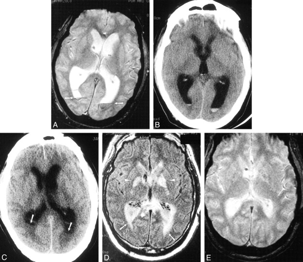
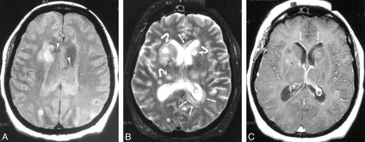
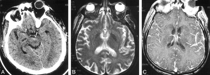
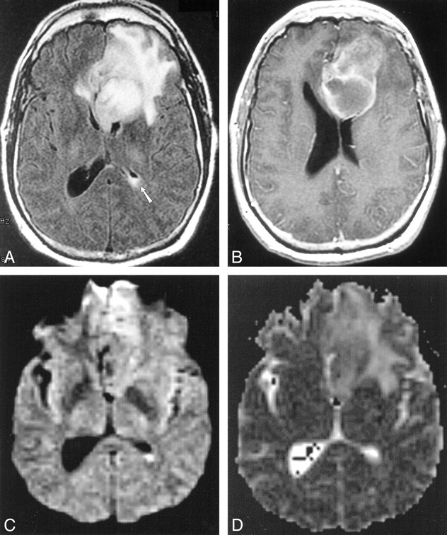
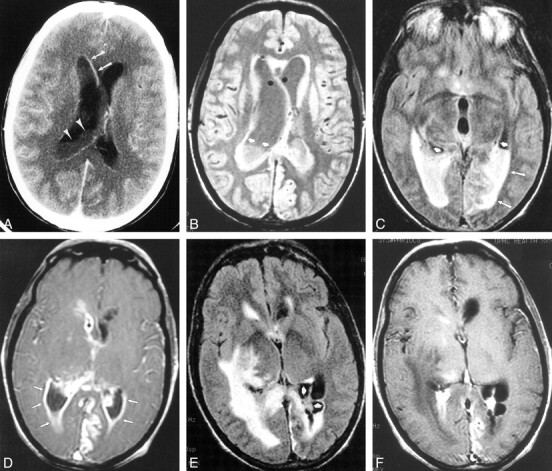
Results: Ventricular debris was detected in 16 (94%) of 17 cases and was irregular in 13 (81%) of 16 cases. Hydrocephalus was present in 13 (76%) of 17 cases. Periventricular hyperintense signal was present in most (seven [78%] of nine) cases with MR imaging and was most conspicuous on fluid-attenuated inversion recovery sequences. Ependymal enhancement was detected in seven (64%) of 11 cases in which contrast material was administered. Signs of meningitis (eg, pial or duraarachnoid signal abnormality or enhancement) were present in 13 (76%) of 17 cases. Three cases had imaging signs of abscess.

Conclusion: Ventricular debris was the most frequent sign of ventriculitis in this series. An irregular level was characteristic of debris in ventriculitis. Hydrocephalus and ependymal enhancement were less frequent signs. Detection of ventricular debris might facilitate diagnosis of pyogenic ventriculitis, a potentially fatal infection, and thus permit appropriate therapy.


Figures

<sc>fig</sc> 1.
fig 1.
Case 3. 41-year-old patient who underwent routine postoperative MR imaging 4 days after resection of a posterior fossa ependymoma. Enterobacter aerogenes grew on culture of ventricular CSF. A, Axial gradient-echo image (950/25/1 [TR/TE/excitation]; flip angle, 20 degrees) shows preexisting ventricular enlargement, related to the tumor, and straight fluid levels with susceptibility artifact within the occipital horns consistent with blood (arrows). B, CT scan from postoperative day 10 also shows this material. C–E, On postoperative day 15, the patient deteriorated. Repeat imaging demonstrates hydrocephalus and irregular debris within the ventricles on CT (C; arrows) and FLAIR images (10,002/145/1 [TR/TEeff/excitation]; inversion time, 2200) (D; open arrows) that does not contain blood products on the gradient-echo image (850/25/1 [TR/TE/excitation]; flip angle, 20 degrees) (E). There also is extensive periventricular hyperintensity (D; arrows), likely representing inflammatory change. Because there was no autopsy, we can only speculate that the hyperintensity in the globus pallidus (arrowheads) might be the result of cerebritis or infarction from vasculitis.
<sc>fig</sc> 2.
fig 2.
Case 13. 58-year-old patient with diabetes presented with altered mental status and multiple abscesses resulting from microaerophilic streptococcus. Gram-positive cocci were found on lumbar CSF analysis as well. Signal abnormality in the periventricular white matter and numerous abscesses (arrows) distributed throughout the deep and subcortical white matter are seen on proton density–weighted (2000/19/1 [TR/TEeff/excitation]) (A) and T2-weighted images (2800/256/1) (B). The irregular ventricular debris (arrowheads) is more clearly demonstrated on the proton density–weighted image (A). T1-weighted image (600/31/1) (C) obtained after intravenous administration of gadopentetate dimeglumine show minimal ependymal enhancement (arrows)
<sc>fig</sc> 3.
fig 3.
Case 12. 66-year-old patient with diabetes and secondary biliary cirrhosis died following infection with E coli meningitis, diagnosed by C1-C2 puncture. Delayed diagnosis occurred as a result of mild signs and symptoms of infection. A, Initial CT scan shows debris and slight hyperattenuation (arrow) compared with CSF, producing an irregular level in the interpeduncular cistern. B, MR imaging was deferred for 2 days because of minor central nervous system symptoms. T2-weighted image (2300/96/2 [TR/TE/excitations]) shows irregular debris (arrows) layering in the lateral ventricles. C, T1-weighted contrast-enhanced image (500/26/1) shows striking pial subarachnoid space enhancement (arrows), but no ependymal enhancement.
<sc>fig</sc> 4.
fig 4.
Case 15. Five months after left frontal craniotomy and resection of an oligodendroglioma, 45-year-old patient presented with headache. Postoperative therapy included adjuvant radiation therapy and steroids for headaches. This MR imaging study prompted a repeat operation in which two abscesses containing gram-positive cocci, one superficial and one deep, were discovered. A, FLAIR image (9002/165/1 [TR/TEeff/excitation]; inversion time, 2200) shows a large left frontal mass with surrounding signal abnormality (abscess at operation) and debris in the left atrium. B, Axial T1-weighted image (500/20/1 [TR/TE/excitation]) shows a cavitary left frontal lesion with peripheral enhancement. At second operation, this proved to be a deep abscess that was in continuity with the ventricle C, Diffusion-weighted image (10,000/96.8/1000 [TR/TEeff/TI]) shows hyperintense signal in the left ventricular debris. D, Apparent diffusion coefficient maps do not show restricted diffusion in the left ventricular pus.
<sc>fig</sc> 5.
fig 5.
Case 14. 42-year-old patient with diabetes initially presented to an outside facility with meningitis and underwent serial imaging demonstrating the imaging course of ventriculitis during a 7-week period. Streptococcus viridans grew on CSF culture. A, Initial contrast-enhanced CT scan shows irregular ventricular debris (arrowheads), hydrocephalus, and ependymal enhancement (arrows). B–D, MR imaging performed 2 weeks later shows ventricular debris (short arrows) and periventricular signal abnormality (long arrows) on proton density–weighted (2000/15/1 [TR/TEeff/excitation]) (B) and FLAIR images (9002/147/1; inversion time, 2200) (C). T1-weighted image (800/20/1) obtained after gadopentetate dimeglumine administration shows extensive ependymal enhancement (arrows) and enhancement of a left posterior cerebral territory infarct. E and F, There is progression of findings on MR imaging performed 7 weeks after initial presentation. FLAIR (10002/155/1; inversion time, 2200) (E) and T1-weighted images (800/8/1) (F) show loculation of ventricles (arrows) with persistent periventricular signal abnormality, but diminished ependymal enhancement.

References

    1. Bakshi R, Kinkel P, Mechtler L, et al. Cerebral ventricular empyema associated with severe adult pyogenic meningitis: computed tomography findings. Clin Neurol Neurosurg 1997;99:252-255 - PubMed
    1. Barloon TJ, Yuh WT, Knepper LE, et al. Cerebral ventriculitis: MR findings. J Comput Assist Tomogr 1990;14:272-275 - PubMed
    1. Bodino J, Lylyk P, Del Valle M, et al. Computed tomography in purulent meningitis. Am J Dis Child 1982;136:495-501 - PubMed
    1. Wormser G, Strashun A. Ventriculitis complicating gram negative meninigitis in an adult: diagnosis by radioisotope brain scanning and computerized tomography. Mt Sinai J Med 1980;47:575-578 - PubMed
    1. Vachon L, Mikity V. Computed tomography and ultrasound in purulent ventriculitis. J Ultrasound Med 1987;6:269-271 - PubMed