

Case Reports
MR imaging and histologic features of capillary telangiectasia of the basal ganglia
Affiliations
- PMID: 11559504
- PMCID: PMC7974575
Item in Clipboard
Case Reports
MR imaging and histologic features of capillary telangiectasia of the basal ganglia
AJNR Am J Neuroradiol.
2001 Sep.
Abstract
Capillary telangiectasias are being recognized with increasing frequency on MR imaging studies. Most are located in the brain stem and show slightly increased signal intensity on T2-weighted images, low signal intensity on T2*-weighted images (reflecting the presence of deoxyhemoglobin), and contrast enhancement. These findings are considered fairly typical for capillary telangiectasia, and pathologic correlation is not generally pursued. We present a case of a proven capillary telangiectasia in the basal ganglia. The imaging features of the lesion were identical to those described for capillary telangiectasias in the brain stem.
Figures
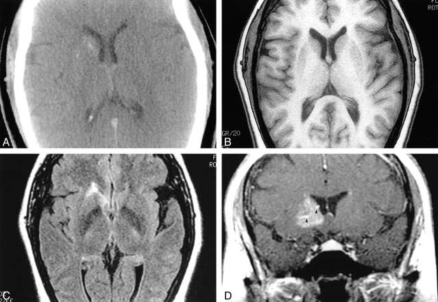
Capillary telangiectasia. A, Noncontrast CT shows slight hyperattenuation in the right basal ganglia with puntacte calcifications. B, Noncontrast MR T1-weighted image is normal. C, Axial FLAIR image (slightly below B) shows high signal intensity in the head of the right caudate nucleus. D, After gadopentate dimeglumine administration, a coronal T1- weighted image shows diffuse enhancement in the right basal ganglia anteriorly. There is a suggestion of large veins (arrowheads) in the malformation.
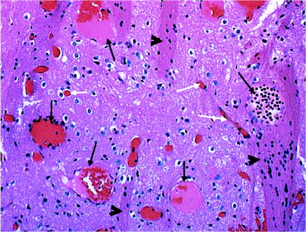
Microscopy. Medium power view (10×). Specimen shows gray matter containing several large, thin-walled vascular spaces (long black arrows). Note normal size capillaries (white arrows). The presence of “pencil fibers” (arrowheads) confirms localization to the basal ganglia. Pencil fibers are the white matter tracts of the basal ganglia. They are an integral part of the histologic composition of the striatum, versus the thalamus, where the neurons are scattered throughout a meshwork of white matter. There is no hemorrhage, hemosiderin-laden macrophages, calcifications, or gliosis
References
-
- Lee RR, Becher MW, Benson ML, Rigamonti D. Brain capillary telangiectasia: MR imaging appearance and clinicohistopathologic findings. Radiology 1997;205:797-805 - PubMed
-
- Auffray-Calvier E, Desal HA, Freund P, Laplaud D, Mathon G, de Kersaint-Gilly A. Capillary telangiectasias: angiographically occult vascular malformations—MRI symptomalogy apropos of 7 cases. J Neuroradiol 1999;26:257-261 - PubMed
-
- Rigamonti D, Johnson PC, Spetzler RF, Hadley MN, Drayer BP. Cavernous malformation and capillary telangiectasia: a spectrum within a single pathological entity. Neurosurgery 1991;28:60-64 - PubMed
Publication types
MeSH terms
LinkOut - more resources
Full Text Sources
Medical