MR imaging in comatose survivors of cardiac resuscitation
- PMID: 11559506
- PMCID: PMC7974574
MR imaging in comatose survivors of cardiac resuscitation
Abstract
Background and purpose: The prognosis of comatose survivors is determined by clinical examination. Early laboratory indicators of poor prognosis (such as evoked potentials) have low sensitivity. The role of MR imaging as a confirmatory study was investigated.
Methods: We studied fluid-attenuated inversion recovery (FLAIR) and diffusion-weighted (DW) imaging in 10 patients comatose after cardiac arrest.
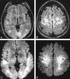
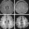
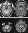
Results: None of the 10 comatose patients had myoclonus status epilepticus or fixed, dilated pupils on neurologic examination, and none had abnormal somatosensory-evoked potentials. Eight patients showed diffuse signal abnormalities, predominantly in the cerebellum (n = 5), the thalamus (n = 8), the frontal and parietal cortices (n = 8), and the hippocampus (n = 9). One patient showed normal MR imaging results, and one patient had abnormalities in the thalamus and cerebellum and minimal abnormality on DW images; both later awakened. None of the patients with abnormal cortical structures on FLAIR MR images recovered beyond a severely disabled state.
Conclusion: MR imaging in comatose survivors may parallel the pathologic findings in severe anoxic-ischemic injury, and extensive abnormalities may indicate little to no prospects for recovery. If confirmed, MR imaging may have a role as a prognosticating test in anoxic-ischemic coma.
Figures


Comment in
-
MR imaging in comatose survivors of cardiac resuscitation.AJNR Am J Neuroradiol. 2002 Apr;23(4):738. AJNR Am J Neuroradiol. 2002. PMID: 11950681 Free PMC article. No abstract available.
References
-
- Levy DE, Caronna JJ, Singer BH, Lapinski RH, Frydman H, Plum F. Predicting outcome from hypoxic-ischemic coma. JAMA 1985;253:1420-1426 - PubMed
-
- Thomassen A, Wernberg M. Prevalence and prognostic significance of coma after cardiac arrest outside intensive care and coronary units. Acta Anaesthesiol Scand 1979;23:143-148 - PubMed
-
- Mullie A, Verstringe P, Buylaert W, et al. Predictive value of Glasgow coma score for awakening after out-of-hospital cardiac arrest. Cerebral Resuscitation Study Group of the Belgian Society for Intensive Care. Lancet 1988;1:137-140 - PubMed
-
- Sandroni C, Barelli A, Piazza O, Proietti R, Mastria D, Boninsegna R. What is the best test to predict outcome after prolonged cardiac arrest? Eur J Emerg Med 1995;2:33-37 - PubMed
-
- Attia J, Cook DJ. Prognosis in anoxic and traumatic coma. Crit Care Clin 1998;14:497-511 - PubMed
MeSH terms
LinkOut - more resources
Full Text Sources
Medical
