. 2001 Sep 15;535(Pt 3):663-77.
doi: 10.1111/j.1469-7793.2001.00663.x.

Gastrin and the neuropeptide PACAP evoke secretion from rat stomach histamine-containing (ECL) cells by stimulating influx of Ca2+ through different Ca2+ channels

Affiliations

Gastrin and the neuropeptide PACAP evoke secretion from rat stomach histamine-containing (ECL) cells by stimulating influx of Ca2+ through different Ca2+ channels

E Lindström et al. J Physiol. .

Abstract

1. Gastrin and PACAP stimulate secretion of histamine and pancreastatin from isolated rat stomach ECL cells. We have examined whether or not secretion depends on the free cytosolic Ca2+ concentration ([Ca2+]i) and the pathways by which gastrin and PACAP elevate [Ca2+]i. Secretion was monitored by radioimmunoassay of pancreastatin and changes in [Ca2+]i by video imaging. The patch clamp technique was used to record whole-cell currents and membrane capacitance (reflecting exocytosis). 2. In the presence of 2 mM extracellular Ca2+, gastrin and PACAP induced secretion and raised [Ca2+]i. Without extracellular Ca2+ (or in the presence of La3+) no secretion occurred. The extracellular Ca2+ concentration required to stimulate secretion was 10 times higher for gastrin than for PACAP. Depletion of intracellular Ca2+ pools by thapsigargin had no effect on the capacity of gastrin and PACAP to stimulate secretion. 3. Gastrin-evoked secretion was inhibited 60-80 % by L-type channel blockers and 40 % by the N-type channel blocker omega-conotoxin GVIA. Combining L-type and N-type channel blockers did not result in greater inhibition than L-type channel blockers alone. Whole-cell patch clamp measurements confirmed that the ECL cells are equipped with voltage-dependent inward Ca2+ currents. A 500 ms depolarising pulse from -60 mV to +10 mV which maximally opened these channels resulted in an increase in membrane capacitance of 100 fF reflecting exocytosis of secretory vesicles. 4. PACAP-evoked secretion was reduced 40 % by L-type channel blockers but was not influenced by inhibition of N-type channels. SKF 96365, a blocker of both L-type and receptor-operated Ca2+ channels, inhibited PACAP-evoked secretion by 85 %. Combining L-type channel blockade with SKF 96365 abolished PACAP-evoked secretion. 5. The results indicate that gastrin- and PACAP-evoked secretion depends on Ca2+ entry and not on mobilisation of intracellular Ca2+. While gastrin stimulates secretion via voltage-dependent L-type and N-type Ca2+ channels, PACAP acts via L-type and receptor-operated Ca2+ channels.


Figures

Figure 1
Figure 1. Increased cytosolic Ca2+ in ECL cells upon PACAP and gastrin stimulation
Stimulation with 10 nm gastrin (A) or PACAP (B) for 3 min in a Ca2+-free medium resulted in a transient increase in [Ca2+]i. The peak rapidly returned back to basal levels. Stimulation with 10 nm gastrin (A) or PACAP (B) for 3 min in a Ca2+-containing medium (1.28 mm CaCl2) resulted in sustained increases in [Ca2+]i.
Figure 2
Figure 2. Increase in membrane capacitance upon infusion of Ca2+-EGTA buffers
A, infusion of a Ca2+-EGTA buffer containing 1.5 μm free Ca2+ or zero (< 1 nm) Ca2+ into single ECL cells caused an increase in membrane capacitance of 12 fF and 0.1 fF s−1, respectively. B, the exocytotic rate as a function of [Ca2+]i. The data points are means of 7, 3, 3 and 12 experiments, respectively, performed with < 1 nm, 160 nm, 365 nm and 1.5 μm free Ca2+ in the pipette solution.
Figure 3
Figure 3. Effect of the extracellular Ca2+ concentration on PACAP- and gastrin-induced pancreastatin secretion
Concentration-reponse curves for gastrin (A) and PACAP (B) in the presence of different extracellular Ca2+ concentrations on pancreastatin secretion. Histograms demonstrating the effect of the extracellular Ca2+ concentration on the maximal pancreastatin secretion induced by 10 nm gastrin (C) or 10 nm PACAP (D). Means ±s.e.m.; n = 8-10.
Figure 4
Figure 4. Effect of thapsigargin on pancreastatin secretion from the ECL cells
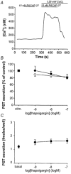
A, ECL cells were pretreated with 100 nm thapsigargin for 4 h. Stimulation with 10 nm PACAP in a Ca2+-free extracellular media did not evoke an increase in [Ca2+]i; however, an increase was evident in Ca2+-containing media. B, concentration-response curve illustrating the lack of effect of pretreatment (4 h) with increasing concentrations of thapsigargin on stimulated pancreastatin (PST) secretion. •, gastrin-induced secretion, ○, PACAP-induced secretion. ▪, PACAP- and gastrin-evoked PST secretion (stim) from controls (set to 100 %). Means ±s.e.m.; n = 6-7.C, effect of thapsigargin per se on pancreastatin secretion from ECL cells. Basal, indicates basal secretion. Means ±s.e.m.; n = 10.
Figure 5
Figure 5. Effect of Ca2+ entry blockers on stimulated pancreastatin secretion from ECL cells
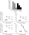
A, histogram illustrating the effect of LaCl3 (La) on both gastrin- and PACAP-induced pancreastatin (PST) secretion. Concentration-response curves demonstrating the effect of the L-type Ca2+ channel blockers nifedipine (B) and nimodipine (C) and the N-type Ca2+ channel blocker ω-conotoxin GVIA (D) on gastrin-induced (•) and PACAP-induced (○) pancreastatin (PST) secretion. ▴, PACAP- and gastrin-evoked secretion (Stim) from controls (secretion set at 100 %). E, concentration-response curve demonstrating the effect of the receptor-operated and L-type channel blocker SKF 96365 on gastrin-induced (•) and PACAP-induced (○) pancreastatin (PST) secretion. ▴, PACAP- and gastrin-evoked secretion (Stim) from controls (secretion set at 100 %). Means ±s.e.m.; n = 6-10.
Figure 6
Figure 6. Effect of combining Ca2+ entry blockers on stimulated pancreastatin secretion from ECL cells
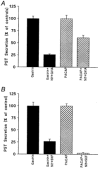
A, effect of combining 10 μm nifedipine (Nif) and 1 μmω-conotoxin GVIA (GVIA) on gastrin- and PACAP-stimulated pancreastatin (PST) secretion. B, effect of combining 10 μm nifedipine and 30 μm SKF 96365 (SKF) on gastrin- and PACAP-stimulated pancreastatin (PST) secretion. Means ±s.e.m.; n = 6-8.
Figure 7
Figure 7. Effect of the L-type Ca2+ channel activator Bay K 8644 on pancreastatin secretion from ECL cell
Concentration-response curve demonstrating the stimulatory effect of the L-type Ca2+ channel activator Bay K 8644 on pancreastatin (PST) secretion. ○, basal secretion. ▴, effect of combining 3 μm Bay K 8644 with 10 μm nifedipine. Means ±s.e.m.; n = 6.
Figure 8
Figure 8. Electrophysiological measurements of Ca2+ currents in single ECL cells
A, ECL cells were depolarised from -60 mV to voltages between -40 and +30 mV in 10 mV increments. For clarity only the responses up to +10 mV are shown. The experiments were performed with 2.6 mm extracellular Ca2+ and in the presence of 0.1 μg ml−1 TTX. B, peak current (I) - voltage (V) relationship for the Ca2+ current. The curve is drawn according to eqn (1). C (left panel), to investigate the presence of L-type and N-type voltage-dependent Ca2+ channels on single ECL cells, inward currents were elicited by 200 ms voltage-clamp depolarisations from -60 to +10 mV and the Ca2+ channel blockers ω-conotoxin GVIA, nifidipine and Cd2+ were added after each other at concentrations of 1 μm, 25 μm and 200 μm, respectively. C (right panel), histogram demonstrating the effect of Ca2+ channel blockers on the peak current. Means ±s.e.m. of 3-8 experiments. D, increase in membrane capacitance upon a 500 ms depolarisation from -60 to 0 mV measured on a single ECL cell in the presence of NaCl.
Figure 9
Figure 9. Electrophysiological measurements of Na+ currents in single ECL cells
A, inward currents from a single ECL cell were elicited by depolarisation from -60 mV to varying voltages between -40 and +10 mV. B, the Na+ peak (the fast transient part of the current) was blocked by application of 0.1 μg ml−1 TTX. C, peak I -V relationship for the Na+ current. The curve was drawn according to eqn (1). Means ±s.e.m. of 6-8 experiments.
Figure 10
Figure 10. Effects of extracellular Na+ on secretion from ECL cells
A, increase in membrane capacitance upon a 500 ms depolarisation from -60 to 0 mV measured on a single ECL cell in the presence of choline chloride (ChCl). B, the mean increase in membrane capacitance in the presence of NaCl or ChCl, respectively. C, histogram showing that 30 min pretreatment with 0.1 μg ml−1 TTX did not affect PACAP- or gastrin-evoked pancreastatin secretion. D, replacing extracellular NaCl with ChCl had no effect on PACAP- or gastrin-evoked pancreastatin secretion. Means ±s.e.m. of 6-8 experiments.

References

    1. Barritt GJ. Receptor-activated Ca2+ inflow in animal cells: a variety of pathways tailored to meet different intracellular Ca2+ signalling requirements. Biochemical Journal. 1999;337:153–169. - PMC - PubMed
    1. Berridge MJ. Elementary and global aspects of calcium signalling. Journal of Physiology. 1997;499:291–306. - PMC - PubMed
    1. Berridge MJ, Irvine RF. Inositol trisphosphate: a novel second messenger in cellular signal transduction. Nature. 1984;312:315–321. - PubMed
    1. Bufler J, Choi GC, Francke C, Schepp W, Prinz C. Voltage-gated Ca2+ currents in rat gastric enterochromaffin-like cells. American Journal of Physiology. 1998;274:C424–429. - PubMed
    1. Burgoyne RD. Control of exocytosis in adrenal chromaffin cells. Biochimica et Biophysica Acta. 1991;1071:174–202. - PubMed

Publication types

MeSH terms

LinkOut - more resources