. 2001 Sep 15;535(Pt 3):679-87.
doi: 10.1111/j.1469-7793.2001.t01-1-00679.x.

Calmodulin kinase and a calmodulin-binding 'IQ' domain facilitate L-type Ca2+ current in rabbit ventricular myocytes by a common mechanism

Affiliations

Calmodulin kinase and a calmodulin-binding 'IQ' domain facilitate L-type Ca2+ current in rabbit ventricular myocytes by a common mechanism

Y Wu et al. J Physiol. .

Retraction in

Abstract

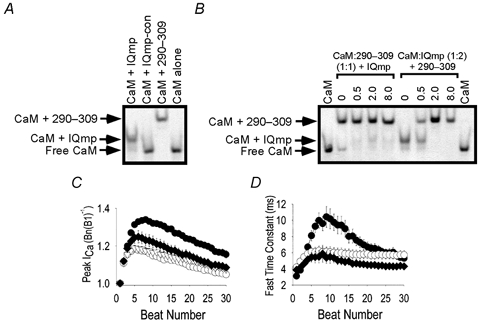
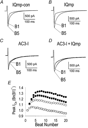
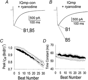
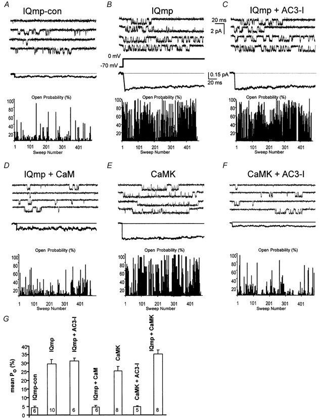
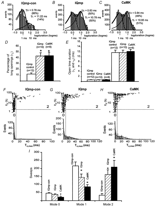
1. Ca2+-calmodulin-dependent protein kinase II (CaMK) and a calmodulin (CaM)-binding 'IQ' domain (IQ) are both implicated in Ca2+-dependent regulation of L-type Ca2+ current (I(Ca)). We used an IQ-mimetic peptide (IQmp), under conditions in which CaMK activity was controlled, to test the relationship between these CaM-activated signalling elements in the regulation of L-type Ca2+ channels (LTCCs) and I(Ca) in rabbit ventricular myocytes. 2. A specific CaMK inhibitory peptide nearly abolished I(Ca) facilitation, but the facilitation was 'rescued' by cell dialysis with IQmp. 3. IQmp significantly enhanced I(Ca) facilitation and slowed the fast component of I(Ca) inactivation, compared with an inactive control peptide. Neither effect could be elicited by a more avid CaM-binding peptide, suggesting that generalized CaM buffering did not account for the effects of IQmp. 4. I(Ca) facilitation was abolished and the fast component of inactivation eliminated by ryanodine, caffeine or thapsigargin, suggesting that the sarcoplasmic reticulum (SR) is an important source of Ca2+ for I(Ca) facilitation and inactivation. IQmp did not restore I(Ca) facilitation under these conditions. 5. Engineered Ca2+-independent CaMK and IQmp each markedly increased LTCC open probability (P(o)) in excised cell membrane patches. The LTCC P(o) increases with CaMK and IQmp were non-additive, suggesting that CaMK and IQmp are components of a shared signalling pathway. 6. Both CaMK and IQmp induced a modal gating shift in LTCCs that favoured prolonged openings, indicating that CaMK and IQmp affect LTCCs through a common biophysical mechanism. 7. These findings support the hypothesis that CaMK is required for physiological I(Ca) facilitation in cardiac myocytes. Both CaMK and IQmp were able to induce a modal gating shift in LTCCs, suggesting that each of these signalling elements is important for Ca2+-CaM-dependent LTCC facilitation in cardiac myocytes.


Figures

Figure 2
Figure 2. Effects of IQmp on L-type ICa facilitation are not due to CaM sequestration
A and B show gel shift assays for determining Ca2+-dependent CaM binding to peptide probes (see Methods). A, the (vertical) lanes are labelled to indicate the presence of CaM and added peptide in a ratio of 1:5. IQmp-con was modified from the original sequence to eliminate CaM binding (see Methods). 290-309 is a peptide that contains a ‘conventional’ CaM-binding domain numbered according to the sequence from CaMK, from which it is derived (Payne et al. 1988). The arrows show that IQmp and 290-309 bound CaM in the presence of Ca2+ (5 μm) and slowed CaM migration through the gel, while IQmp-con did not ‘shift’ CaM migration compared with CaM alone (lane 4). B, the (vertical) lanes are labelled to show the molar proportion of added peptide (IQmp in lanes 2-5 and 290-309 in lanes 6-9) to preformed complexes of CaM:290-309 in equimolar proportions (lanes 2-5) and CaM:IQmp in twofold excess of IQmp over CaM (lanes 6-9). CaM alone was run for comparison (lanes 1 and 10). C and D show summary data for ICa facilitation and inactivation. C, peak ICa, normalized to B1 (as in Fig. 1E), for ventricular myocytes dialysed with IQmp-con (○; n = 10), IQmp (•; n = 10), the CaM-binding peptide 290-309 (♦; n = 7) or solution lacking peptide (▿; n = 23). The data for IQmp-con- and IQmp-treated cells are the same as in Fig. 1E. Differences in ICa facilitation were significant (P = 0.047 for beat 3 and P < 0.001 for subsequent beats) between groups. Differences in ICa facilitation were also significant (P < 0.05) between 290-309-treated cells versus IQmp-con-treated cells for beats 10-20. D, the fast time constant of ICa inactivation was significantly slowed by IQmp (P < 0.01) for beats 6-17. Symbols as in C.
Figure 1
Figure 1. CaMK is required for ICa facilitation, but IQmp is able to facilitate L-type Ca2+ current even after CaMK inhibition
A-D show representative ICa recorded using whole-cell mode voltage clamp. A, a ventricular myocyte dialysed with IQmp-con developed increased ICa and slowed inactivation during successive stimulation pulses (i.e. ‘facilitation’). The first (B1) and fifth (B5) ‘beats’ are superimposed for comparison. B, addition of IQmp enhanced ICa facilitation compared with cells treated with IQmp-con. C, ICa facilitation was nearly eliminated by addition of the specific CaMK inhibitory peptide AC3-I. D, IQmp restored ICa facilitation when co-administered with AC3-I. The holding current was 0 pA. E, summary data for peak ICa, expressed as the ratio of the n th (Bn) to the first (B1) stimulated beat (0.5 Hz) plotted against the beat number for cells treated with IQmp-con (○; n = 10), IQmp (•; n = 10), AC3-I (□; n = 19) and AC3-I + IQmp (▪; n = 12).ICa facilitation was significantly increased in all groups compared with AC3-I-treated cells (P < 0.001) from beat 2 onwards.
Figure 3
Figure 3. SR Ca2+ is required for ICa facilitation
A, ryanodine pretreatment slowed ICa inactivation and eliminated ICa facilitation in the presence of IQmp-con. B, failure of IQmp to restore ICa facilitation in a myocyte treated with ryanodine. In A and B, the holding current was 0 pA. C, summary data (represented as in Fig. 1E) for ICa in myocytes pretreated with ryanodine and then dialysed with IQmp-con (○; n = 8) or IQmp (•; n = 6). No significant differences were found between groups. D, pretreatment with ryanodine eliminated the fast time constant of ICa inactivation; the cells were the same as those shown in C.
Figure 4
Figure 4. LTCC Po is non-additively increased by CaMK and IQmp in excised cell membrane patches
A, under control conditions, LTCCs opened infrequently. B, LTCC Po was markedly increased by IQmp under conditions adverse to endogenous CaMK activation (see Methods). C, addition of the CaMK inhibitory peptide AC3-I did not prevent increased Po in response to IQmp, confirming that the effects of IQmp were independent of ongoing CaMK activity. D, CaM-bound IQmp failed to increase LTCC activity. E, addition of an engineered Ca2+-independent form of CaMK (0.9 μm) increased LTCC Po. F, the effect of CaMK was prevented by the CaMK inhibitory peptide AC3-I, as previously reported (Dzhura et al. 2000). A-F all show representative single-channel sweeps (top), with an ensemble-averaged current (255 to 446 traces, middle), and an all-sweep probability diary (bottom) where the bar height represents the percentage of time the channel was open during the 200 ms depolarization, for each experimental condition. Calibration bars apply to all panels. G, mean LTCC Po was not different for 100 nm or 1 μm (not shown) free bath (intracellular) Ca2+ concentrations. IQmp and CaMK both significantly (P < 0.001) increased LTCC Po compared with control (IQmp-con), but these effects were not additive. LTCC Po was not different between IQmp, CaMK and IQmp + CaMK. The number of cell membrane patches studied is indicated for each group.
Figure 5
Figure 5. Quantification of LTCC open times and analysis of modal gating
A-C, histograms with logarithmically binned open time durations (abscissa) plotted against the number of events (ordinate) (Sigworth & Sine, 1987). LTCC open times were best fitted by the sum of two exponentials; the long (τL) and short (τS) time constants from these fits are shown, with the percentage of total openings described by each time constant in parentheses. These data are from the same cell membrane patches as shown in Fig. 4. D, the percentage of time spent in long LTCC openings was significantly increased by IQmp and CaMK compared with IQmp-con (*P < 0.001). E, LTCC open time durations were not changed by IQmp or CaMK compared with IQmp-con. F-H show LTCC Po plotted against maximum LTCC open time (To,max) in each non-blank sweep. The lower histograms are fitted with two Gaussian distributions, and the vertical lines mark the minimum between the long and short LTCC openings. The horizontal lines indicate Po = 2 % and divide high and low Po sweeps. The gating modes are defined by the intersection of the horizontal and vertical lines (Yue et al. 1990) and are indicated by numerals (0-2). Both IQmp (G) and CaMK (H) increased the number of sweeps with prolonged LTCC openings (mode 2 gating) compared with cell membrane patches exposed to IQmp-con (F). I, summary data for the distribution of gating modes showing that both IQmp and CaMK significantly increased mode 2 gating (*P < 0.001) at the expense of mode 1 gating (*P < 0.001). CaMK also significantly reduced the number of sweeps with low-activity mode 0 gating (†P = 0.035).

Comment in

  • Findings of research misconduct.
    [No authors listed] [No authors listed] NIH Guide Grants Contracts (Bethesda). 2014 Dec 5:NOT-OD-15-031. NIH Guide Grants Contracts (Bethesda). 2014. PMID: 25528784 Free PMC article. No abstract available.
  • Findings of Research Misconduct.
    [No authors listed] [No authors listed] Fed Regist. 2014 Nov 25;79(227):70187-70188. Fed Regist. 2014. PMID: 27737250 Free PMC article. No abstract available.

References

    1. Adachi-Akahane S, Cleemann L, Morad M. Cross-signaling between L-type Ca2+ channels and ryanodine receptors in rat ventricular myocytes. Journal of General Physiology. 1996;108:435–454. - PMC - PubMed
    1. Anderson ME, Braun AP, Schulman H, Premack BA. Multifunctional Ca2+/calmodulin-dependent protein kinase mediates Ca2+-induced enhancement of the L-type Ca2+ current in rabbit ventricular myocytes. Circulation Research. 1994;75:854–861. - PubMed
    1. Bers DM, Patton CW, Nuccitelli R. A practical guide to the preparation of Ca2+ buffers. Methods in Cell Biology. 1994;40:3–29. - PubMed
    1. Braun AP, Schulman H. A non-selective cation current activated via the multifunctional Ca2+-calmodulin-dependent protein kinase in human epithelial cells. Journal of Physiology. 1995;488:37–55. - PMC - PubMed
    1. Chao SH, Suzuki Y, Zysk JR, Cheung WY. Activation of calmodulin by various metal cations as a function of ionic radius. Molecular Pharmacology. 1984;26:75–82. - PubMed

Publication types

Substances

LinkOut - more resources