. 2001 Sep 15;535(Pt 3):825-39.
doi: 10.1111/j.1469-7793.2001.t01-1-00825.x.

Nitric oxide is required for the induction and heterosynaptic spread of long-term potentiation in rat cerebellar slices

Affiliations

Nitric oxide is required for the induction and heterosynaptic spread of long-term potentiation in rat cerebellar slices

S Jacoby et al. J Physiol. .

Abstract

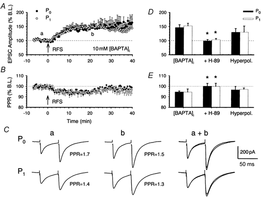
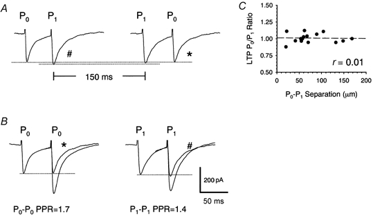
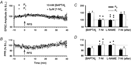
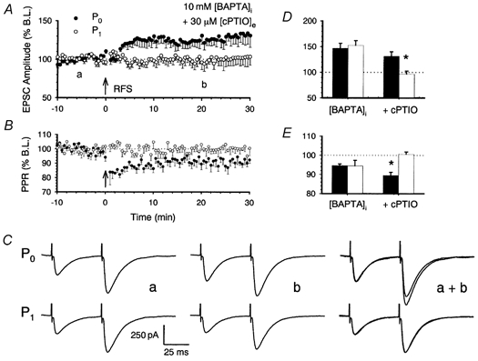
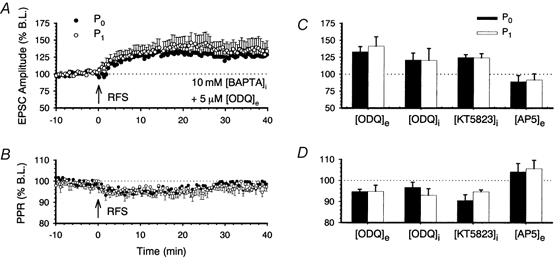
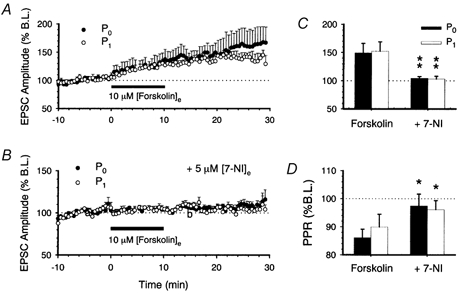
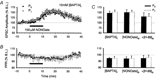
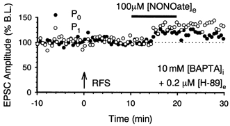
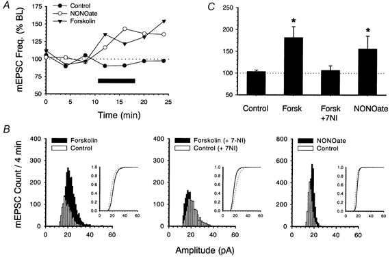
1. In the cerebellar cortex, brief, 8 Hz activation of parallel fibres (PFs) induces a cyclic adenosine 3'5'-monophosphate (cAMP) and protein kinase A (PKA)-dependent form of long-term potentiation between PFs and Purkinje cells. 2. With 10 mM BAPTA in the recording pipette, potentiation evoked by raised frequency stimulation (RFS) to one of two, synaptically independent PF inputs to the same Purkinje cell did not remain input specific but consistently spread to synapses that did not receive RFS, up to the maximum distance tested of 168 microm. 3. LTP at activated and non-activated sites was accompanied by a decrease in paired pulse facilitation (PPF). The PKA inhibitor H-89 blocked both of these effects. Inhibition of nitric oxide synthase (NOS), either by 7-nitro-indazole (7-NI) or N (G)-nitro-L-arginine methyl ester (L-NAME), completely prevented heterosynaptic potentiation and associated reduction in PPF. LTP at distant synapses was selectively prevented by the nitric oxide scavenger 2-(4-carboxyphenyl)-4,4,5,5-tetramethylimidazoline-1-oxyl-3-oxide (cPTIO). Inhibition of soluble guanylate cyclase or protein kinase G had no effect on either pathway. 4. Synaptic potentiation at PF-PC synapses, induced by the adenylate cyclase activator forskolin, was also prevented by inhibition of NOS. Forskolin-induced increases in mEPSC frequency were similarly prevented by NOS inhibition and mimicked by the NO donor spermine NONOate. 5. These results are consistent with the notion that heterosynaptic potentiation is of pre-synaptic origin and dependent upon activation of cAMP/PKA and NO. Moreover, they suggest that cAMP/PKA activation stimulates NO production and this diffusible messenger facilitates pre-synaptic transmitter release at synapses within a radius of upwards of 150 microm, through a mechanism that does not involve cGMP.


Figures

Figure 1
Figure 1. RFS to one of two PF inputs induces PKA-dependent LTP in Purkinje cells that is associated with a decrease in the PPR in both pathways
A, once baseline responses to 0.2 Hz activation of two separate PF inputs to Purkinje cells were stable, one pathway, designated P0 (•), was activated at a raised frequency of 8 Hz for 15 s at time 0 (RFS, arrow). The other pathway (P1; ○) was not activated during this period. Stimulation to both pathways was then resumed at 0.2 Hz. With 10 mm BAPTA included in the patch pipette, RFS led to a long-lasting increase in synaptic responses in both pathways. B, LTP was accompanied by a reduction in the paired-pulse ratio in both pathways. Data are expressed as the percentage change in the ratio of the second pulse to the first compared to baseline levels. C, a single, representative example of EPSCs elicited by stimulation of P0 (top) and P1 (bottom) at times a and b indicated in panel A. The amplitudes of the first pulse in P0 and P1 were normalized to illustrate the accompanying reduction in the PPR (a + b). D, the means and standard errors of P0 (▪) and P1 normalized EPSCs (□) measured 20 min after RFS are shown under conditions of 10 mm intracellular BAPTA, BAPTA plus 0.2 μm extracellular H-89 and following cell hyperpolarization to -90 mV during RFS with 0.5 mm EGTA inside the recording pipette. E, t he associated changes in PPR measured at the same points in time are shown. In graphs (A, B, D and E), the means and s.e.m. of six experiments are shown. Asterisks indicate a statistical difference between test and control conditions (Mann-Whitney U test, P < 0.05).
Figure 2
Figure 2. P0 and P1 display pathway independence
A, parallel fibre pathways P0 and P1 to a single Purkinje cell were activated at 50 ms intervals and again in reverse order 150 ms later. When preceded by activation of the alternate pathway, P0 and P1 EPSC amplitudes remained similar to naïve responses, in this and in four other examples. B, the second response to P1-P0 stimulation (*) and P0-P1 stimulation (#) shown in A are superimposed on P0-P0 and P1-P1 responses, respectively. C, the ratios of the percentage level of potentiation compared to baseline levels observed in P0 compared to that in P1 are plotted against the separations between electrodes for a group of 15 cells. The dashed line represents the fitted linear regression.
Figure 3
Figure 3. RFS-induced LTP requires NOS activity
A, inhibition of NOS with 7-NI in the extracellular perfusate prevented LTP in pathways P0 and P1. B, the associated mean changes in PPR over time are shown. C, a comparison of P0 and P1 responses recorded under standard control conditions of 10 mm intracellular BAPTA and in the presence of 7-NI and l-NAME. Application of 5 μm 7-NI to the bathing medium 5 min after RFS had no effect on the extent of LTP in either P0 or P1. D, the associated changes in PPR are shown. The means and s.e.m. of 6 (7-NI), 5 (l-NAME) and 4 (7-NI application 5 min after RFS) cells are shown. Asterisks indicate a statistical difference between test and control conditions (Mann-Whitney U test, *P < 0.05; **P < 0.01).
Figure 4
Figure 4. RFS induces input-specific LTP in the presence of the NO scavenger cPTIO
With 10 mm BAPTA in the patch pipette and 30 μm cPTIO in the extracellular perfusate, RFS to pathway P0 led to an input-specific, long-lasting potentiation of P0 synaptic responses (A) and an input-specific reduction in the paired pulse ratio (B). Examples of representative P0 and P1 EPSCs taken at times a and b indicated in panel A are shown below (C. D), the means and s.e.m. of P0 (▪) and P1 normalized EPSCs (□) measured 20 min after RFS are shown under control conditions (10 mm intracellular BAPTA) and with cPTIO in the perfusate together with changes in the PPR (E). In graphs (A, B, D and E), the means and s.e.m. of six experiments are shown. Asterisks indicate a statistical difference between P0 and P1 pathways (Wilcoxon signed-rank test, P < 0.01).
Figure 5
Figure 5. RFS-induced LTP does not require guanylate cyclase
Inhibition of guanylate cyclase with 5 μm extracellular ODQ did not prevent LTP (A) or the associated changes in PPR (B) in either P0 or P1. Similarly, neither intracellular ODQ (5 μm) nor the PKG inhibitor KT5823 (500 nm) prevented LTP or the decrease in PPR (D). Potentiation (C) and the reduction in PPR (D) was prevented in both pathways when the NMDA receptor antagonist AP5 (50 μm) was present in the perfusate. In all cases, the means and s.e.m. of six cells are shown.
Figure 6
Figure 6. Forskolin induces LTP
A, 10 min bath application of 10 μm forskolin led to a sustained potentiation of P0 and P1 responses. B, inhibition of NOS with extracellular 7-NI prevented forskolin-induced LTP. C, a comparison of P0 and P1 responses recorded in the presence of forskolin and additionally with 7-NI. Asterisks indicate statistical difference between test and control conditions (Mann-Whitney U test): **P < 0.01, *P < 0.05. D, the associated changes in normalized PPR are illustrated. The means and s.e.m. of six cells are shown.
Figure 7
Figure 7. Application of NO results in LTP
A, 10 min bath application of the NO donor spermine NONOate induced an increase of responses in both pathways. B, illustration of the associated change in PPR over time. C, a comparison of P0 and P1 responses measured 20 min after RFS under control conditions of 10 mm BAPTA and 20 min after NONOate application in the absence and the presence of H-89.
Figure 8
Figure 8. NO donor-induced potentiation does not require PKA
In the presence of 0.2 μm H-89, 15 s RFS (arrow) failed to induce potentiation. Subsequent application of 100 μm spermine NONOate (horizontal bar) produced a potentiation of P0 and P1 responses.
Figure 9
Figure 9. Forskolin increases mEPSC frequency via NOS and NO activities
A, changes in mEPSC frequency over time expressed as a percentage of baseline levels under control conditions of 10 mm intracellular BAPTA. The effects of application of 10 μm forskolin and 100 μm NONOate are shown. B, the frequency distributions of mEPSCs over two 4 min periods, prior to (□) and 15 min after application of forskolin (▪) in absence (left) and presence (centre) of 7-NI and the frequency distributions of mEPSCs recorded prior to and 15 min after application of 10 μm spermine NONOate (right) are shown. Inset are shown cumulative probability curves. C, means and s.e.m. of mEPSC frequency recorded under standard conditions, in the presence of forskolin (Forsk), in the additional presence of 7-NI, and after NONOate application. Asterisks represent a statistical difference (P < 0.01) between control and test conditions (Kolmogorov-Smirnov test).

Similar articles

Cited by

References

    1. Ahn S, Ginty DD, Linden DJ. A late phase of cerebellar long-term depression requires activation of CaMKIV and CREB. Neuron. 1999;23:559–568. - PubMed
    1. Anderson WW, Collingridge GL. A data acquisition program for on-line analysis of long-term potentiation and long-term depression. Society for Neuroscience Abstracts. 1999;23:665. - PubMed
    1. Arancio O, Kiebler M, Lee CJ, Levram V, Tsien RY, Kandel ER, Hawkins RD. Nitric-oxide acts directly in the presynaptic neuron to produce long-term potentiation in cultured hippocampal-neurons. Cell. 1996;87:1025–1035. - PubMed
    1. Ariano MA, Lewicki JA, Brandwein HJ, Murad F. Immunohistochemical localization of guanylate cyclase within neurons of rat brain. Proceedings of the National Academy of Sciences of the USA. 1982;79:1316–1320. - PMC - PubMed
    1. Atluri PP, Regehr WG. Determinants of the time course of facilitation at the granule cell to Purkinje cell synapse. Journal of Neuroscience. 1996;16:5661–5671. - PMC - PubMed

Publication types

MeSH terms

LinkOut - more resources