

Selection for loss of p53 function in T-cell lymphomagenesis is alleviated by Moloney murine leukemia virus infection in myc transgenic mice
- PMID: 11559812
- PMCID: PMC114551
- DOI: 10.1128/JVI.75.20.9790-9798.2001
Selection for loss of p53 function in T-cell lymphomagenesis is alleviated by Moloney murine leukemia virus infection in myc transgenic mice
Abstract
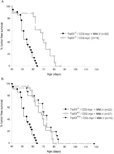
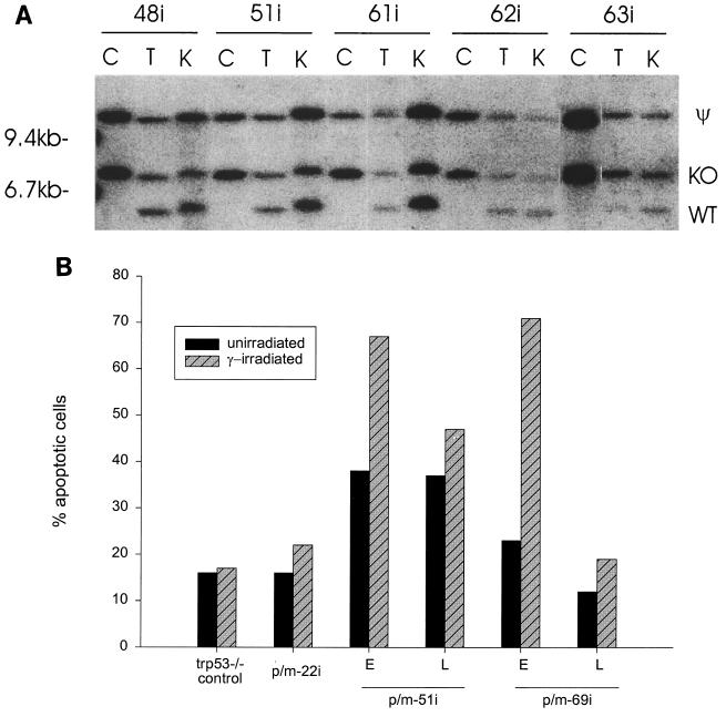
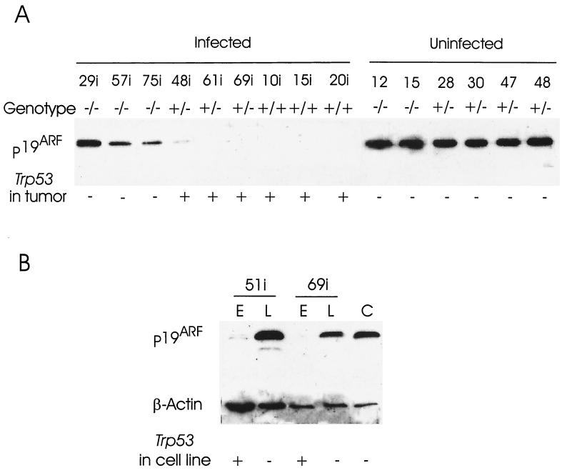
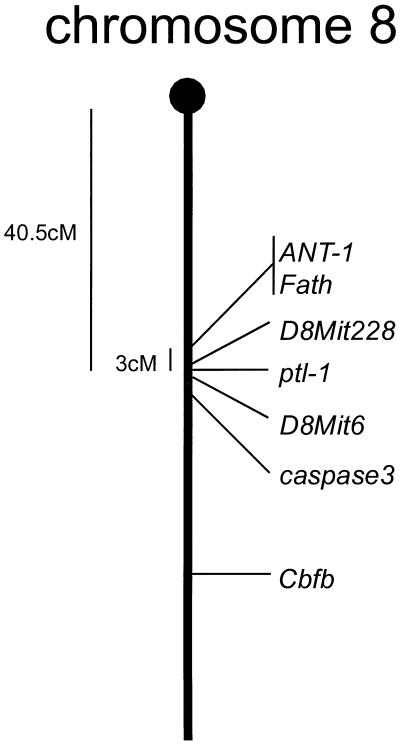
Thymic lymphomas induced by Moloney murine leukemia virus (MMLV) have provided many examples of oncogene activation, but the role of tumor suppressor pathways in these tumors is less clear. These tumors display little evidence of loss of heterozygosity, and MMLV is only weakly synergistic with the Trp53 null genotype, suggesting that viral lymphomagenesis involves mechanisms which do not require mutational loss of Trp53 function. To explore this relationship in greater depth, we infected CD2-myc transgenic mice with MMLV and examined the role of Trp53 in the genesis of these tumors. Most (19 of 27) of the tumors from MMLV-infected, CD2-myc Trp53(+/-) mice retained the wild-type Trp53 allele in vivo while tumors of uninfected CD2-myc Trp53(+/-) mice invariably showed allele loss from a significant fraction of primary tumor cells. The functional integrity of the Trp53 gene in these tumors was indicated by ongoing allele loss or selection for mutational stabilization during in vitro propagation and by the radiosensitivity of selected Trp53(+/-) tumor cell lines. An inverse correlation was noted between retention of the wild-type Trp53 allele and expression of p19(ARF), providing further evidence of negative-feedback control of the latter by p53. However, expression of p19(ARF) does not appear to be counterselected in the absence of p53, and its integrity in Trp53(+/-) tumors was indicated by its transcriptional upregulation on Trp53 wild-type allele loss in vitro in selected tumor cell lines. The role of MMLV was investigated further by analysis of proviral insertion sites in tumors of CD2-myc transgenic mice sorted for Trp53 genotype. A proportion of tumors showed insertions at Runx2, an oncogene which has been shown to collaborate independently with CD2-myc and with the Trp53 null genotype, and at a novel common integration site (ptl-1) on chromosome 8. Genotypic analysis of the panel of tumors suggested that neither of these integrations is functionally redundant with loss of p53, but it appears that the combination of the MMLV oncogenic program with the CD2-myc oncogene relegates p53 loss to a late step in tumor progression or in vitro culture. While the means by which these tumors preempt the p53 tumor suppressor response remains to be established, this study provides further evidence that irreversible inactivation of this pathway is not a prerequisite for tumor development in vivo.
Figures
Similar articles
-
Synergy between a human c-myc transgene and p53 null genotype in murine thymic lymphomas: contrasting effects of homozygous and heterozygous p53 loss.Oncogene. 1995 May 4;10(9):1717-23. Oncogene. 1995. PMID: 7753548
-
Activation of a novel proto-oncogene, Frat1, contributes to progression of mouse T-cell lymphomas.EMBO J. 1997 Feb 3;16(3):441-50. doi: 10.1093/emboj/16.3.441. EMBO J. 1997. PMID: 9034327 Free PMC article.
-
The MMTV/c-myc transgene and p53 null alleles collaborate to induce T-cell lymphomas, but not mammary carcinomas in transgenic mice.Oncogene. 1995 Jul 6;11(1):181-90. Oncogene. 1995. PMID: 7624126
-
Friend virus induced murine erythroleukaemia: the p53 locus.Cancer Surv. 1992;12:137-51. Cancer Surv. 1992. PMID: 1638545 Review.
-
Tumorigenesis in transgenic mice: identification and characterization of synergizing oncogenes.J Cell Biochem. 1991 Oct;47(2):130-5. doi: 10.1002/jcb.240470206. J Cell Biochem. 1991. PMID: 1661736 Review.
Cited by
-
Dosage-dependent tumor suppression by histone deacetylases 1 and 2 through regulation of c-Myc collaborating genes and p53 function.Blood. 2013 Mar 14;121(11):2038-50. doi: 10.1182/blood-2012-08-450916. Epub 2013 Jan 17. Blood. 2013. PMID: 23327920 Free PMC article.
-
The common retroviral insertion locus Dsi1 maps 30 kilobases upstream of the P1 promoter of the murine Runx3/Cbfa3/Aml2 gene.J Virol. 2002 May;76(9):4364-9. doi: 10.1128/jvi.76.9.4364-4369.2002. J Virol. 2002. PMID: 11932403 Free PMC article.
-
Insertional mutagenesis and deep profiling reveals gene hierarchies and a Myc/p53-dependent bottleneck in lymphomagenesis.PLoS Genet. 2014 Feb 27;10(2):e1004167. doi: 10.1371/journal.pgen.1004167. eCollection 2014 Feb. PLoS Genet. 2014. PMID: 24586197 Free PMC article.
-
Long-range effects of retroviral insertion on c-myb: overexpression may be obscured by silencing during tumor growth in vitro.J Virol. 2003 Jan;77(2):1059-68. doi: 10.1128/jvi.77.2.1059-1068.2003. J Virol. 2003. PMID: 12502821 Free PMC article.
-
Decreased virus population diversity in p53-null mice infected with weakly oncogenic Abelson virus.J Virol. 2005 Sep;79(18):11618-26. doi: 10.1128/JVI.79.18.11618-11626.2005. J Virol. 2005. PMID: 16140739 Free PMC article.
References
-
- Blyth K, Stewart M, Bell M, James C, Evan G, Neil J C, Cameron E R. Sensitivity to myc-induced apoptosis is retained in spontaneous and transplanted lymphomas of CD2-mycER mice. Oncogene. 2000;19:773–782. - PubMed
-
- Blyth K, Terry A, Mackay N, Vaillant F, Bell M, Cameron E R, Neil J C, Stewart M. Runx2: a novel oncogenic pathway revealed by in vivo complementation and retroviral tagging. Oncogene. 2001;20:295–302. - PubMed
-
- Blyth K, Terry A, O'Hara M, Baxter E W, Campbell M, Stewart M, Donehower L A, Onions D E, Neil J C, Cameron E R. Synergy between a human c-myc transgene and p53 null genotype in murine thymic lymphomas–contrasting effects of homozygous and heterozygous p53 loss. Oncogene. 1995;10:1717–1723. - PubMed
-
- Crouch D H, Lang C, Gillespie D A H. The leucine zipper motif of avian c-myc is required for transformation and autoregulation. Oncogene. 1990;5:683–689. - PubMed
Publication types
MeSH terms
Substances
Associated data
- Actions
LinkOut - more resources
Full Text Sources
Molecular Biology Databases
Research Materials
Miscellaneous
