. 2001 Oct;75(20):9857-71.
doi: 10.1128/JVI.75.20.9857-9871.2001.

Transient disruption of intercellular junctions enables baculovirus entry into nondividing hepatocytes

Affiliations

Transient disruption of intercellular junctions enables baculovirus entry into nondividing hepatocytes

J P Bilello et al. J Virol. 2001 Oct.

Abstract

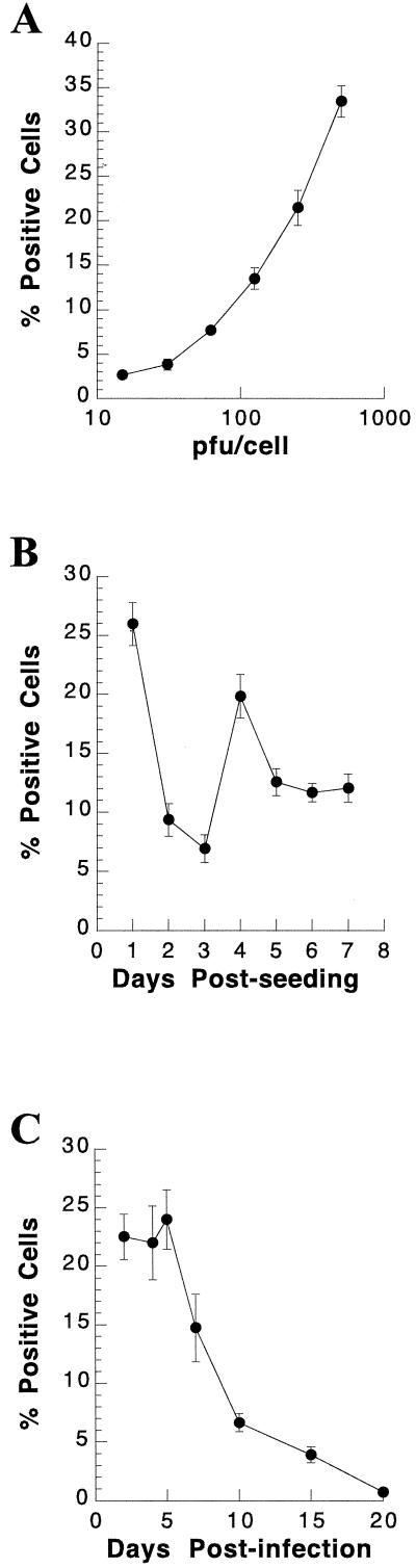
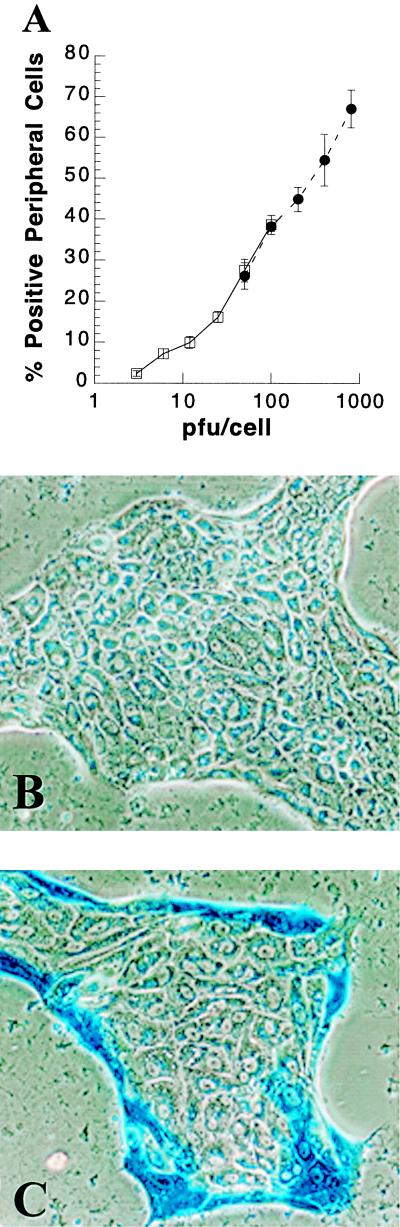
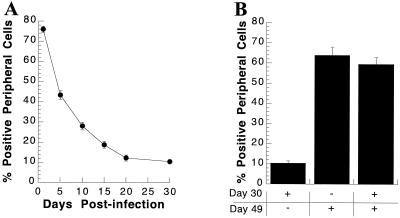
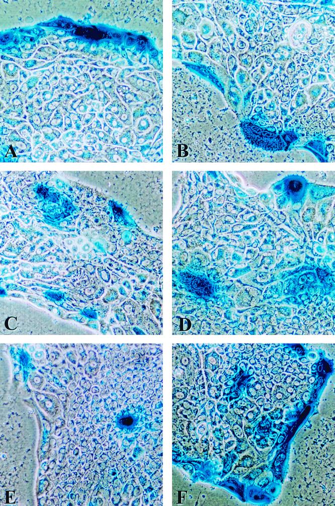
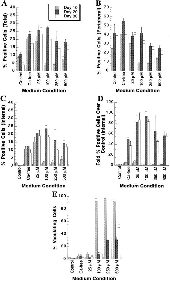
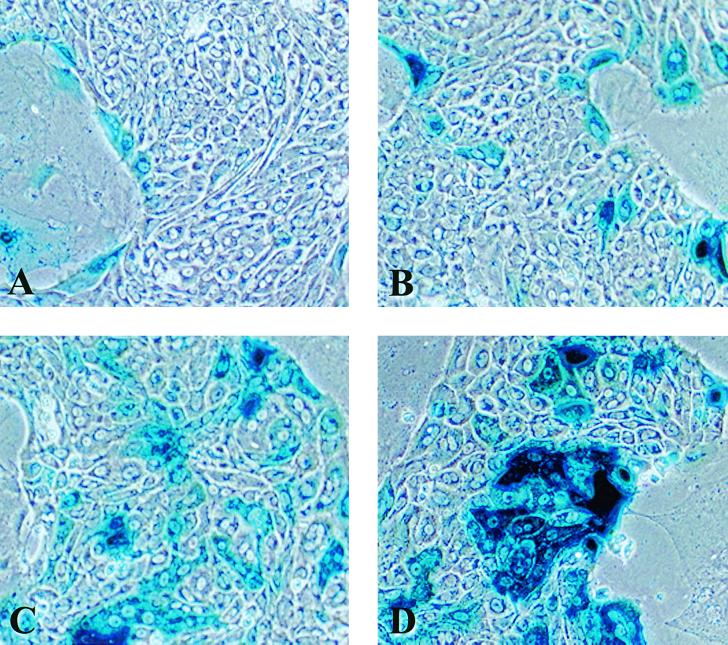
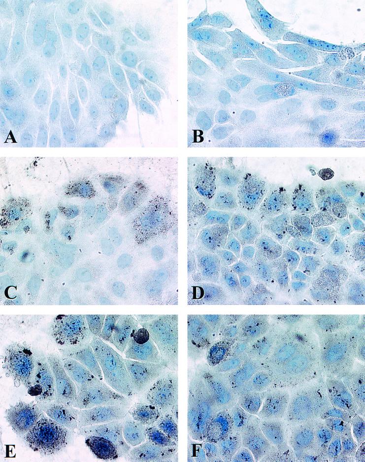
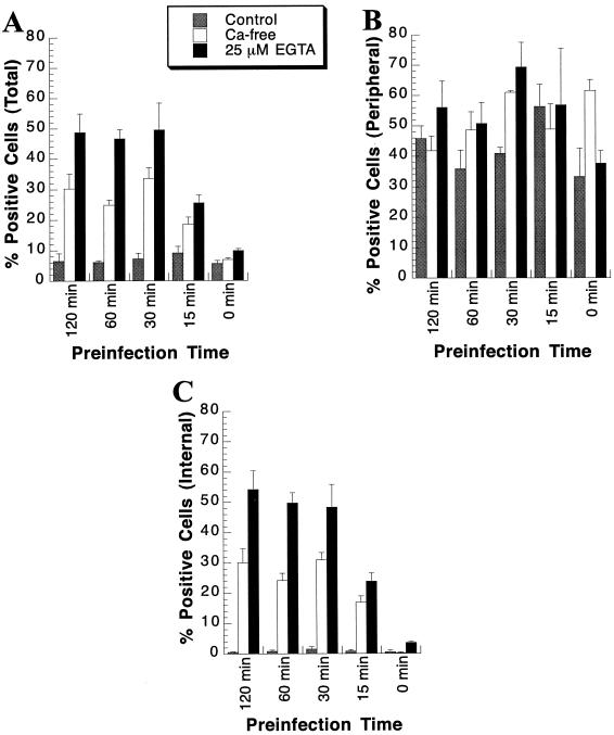
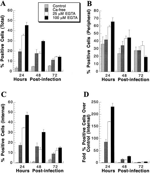
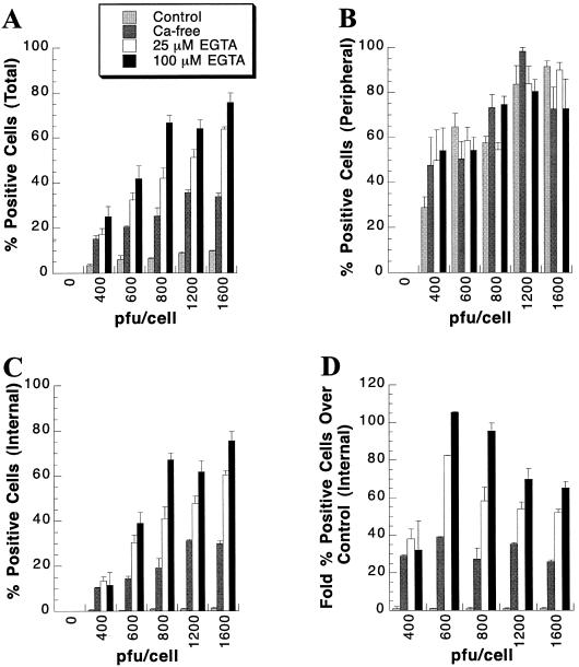
Baculovirus infection has extended the capabilities for transfection of exogenous genes into a variety of mammalian cell types. Because rat hepatocytes plated on collagen-coated dishes and maintained in dimethyl sulfoxide (DMSO)-supplemented chemically defined medium are an excellent model system for studying liver function in vitro, we investigated the ability of baculoviruses to infect and deliver exogenous genes to cells in this culture system. Efficient delivery to hepatocytes in short-term culture becomes restricted to peripheral cells, or "edge" cells, as the hepatocytes acquire intercellular junctions and form islands with time in culture. This barrier to baculovirus entry can be overcome, and the percentage of internal cells within the hepatocyte islands that are infected with the baculovirus can be increased more than 100-fold, when cells are subjected to transient calcium depletion before and during infection. These findings suggest that at least in some cell types, such as hepatocytes, baculovirus entry may require contact with the basolateral surface. We conclude from this study that recombinant baculovirus infection following transient depletion of extracellular calcium results in delivery of exogenous genes to at least 75% of hepatocytes in long-term DMSO culture, thereby making it possible for the first time to carry out gain-of-function and loss-of-function studies in this cell system.


Figures

FIG. 1
FIG. 1
Baculovirus-mediated gene transfer into primary rat hepatocytes in short-term culture. (A) Dose dependence of baculovirus-mediated gene transfer into short-term culture. Primary rat hepatocytes were perfused and seeded on collagen-coated dishes. Cultures were infected with CMV-lacZ baculovirus at the indicated multiplicities 24 h postseeding. At 24 h p.i., primary hepatocyte cultures were stained for β-Gal activity as described in Materials and Methods, and the percent β-Gal-positive cells was determined. The mean percent β-Gal-positive cells, with the standard deviation, is shown as a function of the PFU of CMV-lacZ baculovirus/cell used to infect primary rat hepatocyte cultures. (B) Effect of time in culture on susceptibility of primary rat hepatocytes to baculovirus-mediated gene transfer. Primary rat hepatocytes were perfused, seeded on collagen-coated dishes, and infected with 300 PFU of CMV-lacZ baculovirus/cell on the indicated days postseeding. At 24 h p.i., cultures were stained for β-Gal activity and the percent positive cells was determined. Each symbol represents the mean percentage of primary rat hepatocytes at the indicated day postseeding that were β-Gal positive, with the standard deviation. (C) Detection of β-Gal activity at various times after infection of hepatocytes in short-term culture. Primary rat hepatocytes were seeded on collagen-coated dishes and infected with 200 PFU of CMV-lacZ baculovirus/cell 24 h postseeding. Cultures were stained for β-Gal activity at the indicated number of days p.i., and the percent β-Gal-positive cells was determined. Each symbol represents the mean percentage of primary rat hepatocytes that stained β-Gal positive at the indicated day p.i., with the standard deviation.
FIG. 2
FIG. 2
Dose-dependent baculovirus-mediated gene transfer into primary rat hepatocytes in long-term DMSO culture. (A) Fifty-five days postseeding, primary rat hepatocytes in long-term DMSO culture were infected with 3, 6, 12, 25, 50, or 100 PFU of CMV-lacZ baculovirus/cell. At 24 h p.i., cultures were stained for β-Gal activity and the percent positive peripheral (edge) cells was determined (□). In a separate experiment, primary rat hepatocytes in long-term DMSO culture for 56 days were infected with 50, 100, 200, 400, or 800 PFU of CMV-lacZ baculovirus/cell (●). At 24 h p.i., cultures were stained for β-Gal activity. Results show the percentage of peripheral hepatocytes positive for β-Gal activity, with the standard deviation, at the indicated PFU of CMV-lacZ baculovirus/cell. (B) Primary rat hepatocytes in long-term DMSO culture for 56 days were mock infected and stained for β-Gal activity 24 h later. (C) Primary hepatocytes in long-term DMSO culture for 56 days were infected with 800 PFU of CMV-lacZ baculovirus/cell and stained for β-Gal activity at 24 h p.i.
FIG. 3
FIG. 3
Detection of β-Gal activity at various times after infection of long-term cultures of primary hepatocytes and susceptibility of peripheral cells to reinfection with CMV-lacZ baculovirus. (A) Detection of β-Gal activity at various times after infection of a long-term DMSO culture. Thirty days postseeding, primary rat hepatocytes in DMSO culture were infected with 400 PFU of CMV-lacZ baculovirus/cell. At the indicated time p.i., cultures were stained for β-Gal activity and the percent β-Gal-positive peripheral (edge) cells was determined. The results show the percentage of peripheral cells positive for β-Gal activity at the indicated time p.i., with the standard deviation. Where no error bar is shown, the error falls within the size of the symbol. (B) Susceptibility of peripheral cells to reinfection. At day 30 postseeding, hepatocytes were either mock infected or infected with 400 PFU of CMV-lacZ baculovirus/cell. Cultures were divided into three groups. One group of cultures infected on day 30 was mock infected at day 49 (+, −), one group of cultures infected at day 30 was reinfected at day 49 (+, +), and the cultures mock infected at day 30 were infected at day 49 (−, +). All cultures were fixed and stained for β-Gal activity at 50 days postseeding. Each bar represents the mean percentage of peripheral (edge) hepatocytes positive for β-Gal activity, with the standard deviation, following infection at the indicated day postseeding.
FIG. 4
FIG. 4
Motility of primary hepatocytes in long-term DMSO culture. Primary rat hepatocytes in long-term DMSO culture for 30 days were infected with 400 PFU of CMV-lacZ baculovirus/cell. Cultures were stained for β-Gal activity after 1 (A), 5 (B), 10 (C), 15 (D), and 30 (E) days p.i. (F) Susceptibility to reinfection of peripheral (edge) cells in long-term primary rat hepatocyte cultures maintained in DMSO. A culture infected on day 30 postseeding was reinfected with an additional 400 PFU of CMV-lacZ baculovirus/cell on day 49 and was stained for β-Gal activity at 24 h p.i.
FIG. 5
FIG. 5
Effect of calcium depletion on baculovirus-mediated gene delivery of CMV-lacZ to primary rat hepatocyte cultures. At the indicated days postseeding, primary rat hepatocytes were pretreated in either control medium (DMEM–F12; designated Control), calcium-free DMEM (Ca-free), or calcium-free DMEM supplemented with either 25 (25 μM), 100 (100 μM), 250 (250 μM), or 500 (500 μM) μM EGTA for 1 h. Subsequently, cultures were infected for 1 h with 400 PFU of CMV-lacZ baculovirus/cell diluted in either control medium, calcium-free DMEM, or calcium-free DMEM supplemented with either 25, 100, 250, or 500 μM EGTA. At 24 h p.i., cultures were stained for β-Gal activity. Percent β-Gal-positive cells was determined for the total culture (A), the peripheral cells (B), and the internal cells (C). (D) Fold increase in internal β-Gal-positive cells following extracellular calcium depletion relative to the control medium. Each bar represents the mean percent β-Gal-positive primary rat hepatocytes, with the standard deviation, at the indicated medium condition. (E) Percent cytopathic cells present in the primary rat hepatocyte cultures following calcium depletion prior to and during baculovirus infection. Each bar represents the mean percent cytopathic cells at the indicated medium condition, with the standard deviation.
FIG. 6
FIG. 6
Effect of calcium depletion on baculovirus-mediated gene delivery to internal hepatocytes. At day 20 postseeding, primary rat hepatocytes were pretreated in either control medium, calcium-free DMEM, or calcium-free DMEM supplemented with either 25 or 100 μM EGTA for 1 h. Subsequently, cultures were infected for 1 h with 400 PFU of CMV-lacZ baculovirus/cell diluted in either control medium (A), calcium-free DMEM (B), or calcium-free DMEM supplemented with either 25 (C) or 100 (D) μM EGTA. At 24 h p.i., cultures were stained for β-Gal activity.
FIG. 7
FIG. 7
Immunoperoxidase staining for AcMNPV in primary rat hepatocytes. Twenty-one days postseeding, primary rat hepatocytes were pretreated in either control medium, calcium-free DMEM, or calcium-free DMEM supplemented with either 25 or 100 μM EGTA for 1 h. Subsequently, cultures were infected for 1 h with 400 PFU of CMV-lacZ baculovirus/cell diluted in either control medium, calcium-free DMEM, or calcium-free DMEM supplemented with either 25 or 100 μM EGTA. At 3 h p.i., cultures were fixed and incubated with an antibody to AcMNPV (A, C, D, E, and F) or a control antibody (B) as described in Materials and Methods. (A) Primary rat hepatocytes at 21 days postseeding that were pretreated and mock infected in control medium. (B) Cultures pretreated and infected with the CMV-lacZ baculovirus in control medium and incubated with a rabbit IgG control antibody. (C) Cultures pretreated and infected in control medium. (D) Cultures pretreated and infected in calcium-free DMEM. (E and F) Cultures pretreated and infected under calcium depletion conditions of 25 and 100 μM, respectively.
FIG. 8
FIG. 8
Determination of the proper pretreatment time prior to infection affording the maximal baculovirus-mediated gene delivery efficiency. At 20 days postseeding, primary rat hepatocytes were pretreated for the indicated times in either control medium (shaded bars), calcium-free DMEM (open bars), or calcium-free DMEM supplemented with 25 μM EGTA (solid bars). Subsequently, cultures were infected for 1 h with 400 PFU of CMV-lacZ baculovirus/cell diluted in either control medium, calcium-free DMEM, or calcium-free DMEM supplemented with 25 μM EGTA. At 24 h p.i., cultures were stained for β-Gal activity. The percent β-Gal-positive cells was determined for the total culture (A), the peripheral cells (B), and the internal cells (C). Each bar represents the mean percent β-Gal-positive primary rat hepatocytes, with the standard deviation, at the indicated pretreatment time prior to CMV-lacZ baculovirus infection.
FIG. 9
FIG. 9
Detection of β-Gal activity at various times after infection of long-term cultures of primary hepatocytes following calcium depletion. Twenty days postseeding, primary rat hepatocytes were pretreated in either control medium (light shaded bars), calcium-free DMEM (dark shaded bars), or calcium-free DMEM supplemented with either 25 (open bars) or 100 (solid bars) μM EGTA for 1 h. Subsequently, cultures were infected for 1 h with 400 PFU of CMV-lacZ baculovirus/cell diluted in either control medium, calcium-free DMEM, or calcium-free DMEM supplemented with either 25 or 100 μM EGTA. At the indicated times p.i., cultures were stained for β-Gal activity. The percent β-Gal-positive cells was determined for the total culture (A), the peripheral cells (B), and the internal cells (C). (D) Fold increase in internal β-Gal-positive cells following extracellular calcium depletion relative to β-Gal-positive internal cells in the control medium. Each bar represents the mean percent β-Gal-positive primary rat hepatocytes at the indicated time p.i., with the standard deviation.
FIG. 10
FIG. 10
Increased dose-dependent gene transfer into primary rat hepatocytes under calcium-depleted conditions. Twenty-one days postseeding, primary rat hepatocytes were pretreated in either control medium (light shaded bars), calcium-free DMEM (dark shaded bars), or calcium-free DMEM supplemented with either 25 (open bars) or 100 (solid bars) μM EGTA for 1 h. Subsequently, cultures were infected for 1 h with the indicated MOI of CMV-lacZ baculovirus/cell diluted in either control medium, calcium-free DMEM, or calcium-free DMEM supplemented with either 25 or 100 μM EGTA. At 24 h p.i., cultures were stained for β-Gal activity. The percent β-Gal-positive cells was determined for the total culture (A), the peripheral cells (B), and the internal cells (C). (D) Fold increase in internal β-Gal-positive cells following extracellular calcium depletion over the level in control medium. Each bar represents the mean percent β-Gal-positive primary rat hepatocytes at the indicated PFU of CMV-lacZ baculovirus/cell, with the standard deviation.

Similar articles

Cited by

References

    1. Bhat M, Toledo-Velasquez D, Wang L, Malanga C J, Ma J K H, Rojanasakul Y. Regulation of tight junction permeability by calcium mediators and cell cytoskeleton in rabbit tracheal epithelium. Pharm Res. 1993;10:991–997. - PubMed
    1. Borrmann C M, Mertens C, Schmidt A, Langbein L, Kuhn C, Franke W W. Molecular diversity of plaques of epithelial-adhering junctions. Ann N Y Acad Sci. 2000;915:144–150. - PubMed
    1. Bour E S, Ward L K, Cornman G A, Isom H C. Tumor necrosis factor-alpha-induced apoptosis in hepatocytes in long-term culture. Am J Pathol. 1996;148:485–495. - PMC - PubMed
    1. Boyce F M, Bucher N L. Baculovirus-mediated gene transfer into mammalian cells. Proc Natl Acad Sci USA. 1996;93:2348–2352. - PMC - PubMed
    1. Boyce F M, Franco E A. High-efficiency transduction of mammalian cells using baculovirus: practical aspects. In: Cid-Arrequi A, Garcia-Carranca A, editors. Viral vectors: basic science and gene therapy. Natick, Mass: Eaton Publishing; 2000. pp. 359–367.

Publication types

LinkOut - more resources