. 2001 Oct;75(20):10005-13.
doi: 10.1128/JVI.75.20.10005-10013.2001.

Differentiation-dependent chromatin rearrangement coincides with activation of human papillomavirus type 31 late gene expression

Affiliations

Differentiation-dependent chromatin rearrangement coincides with activation of human papillomavirus type 31 late gene expression

L M del Mar Peña et al. J Virol. 2001 Oct.

Abstract

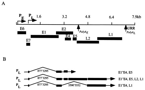
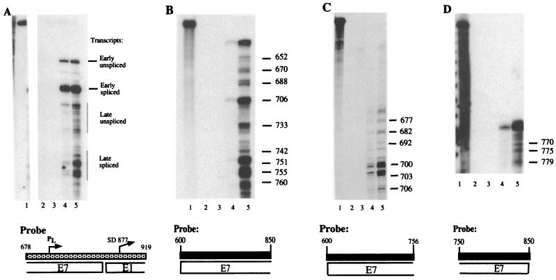
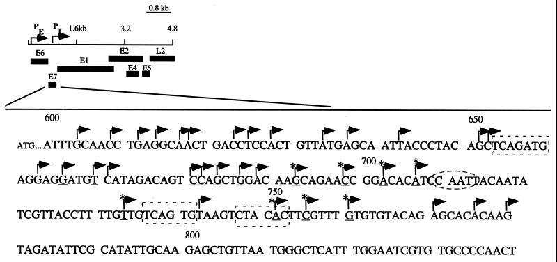
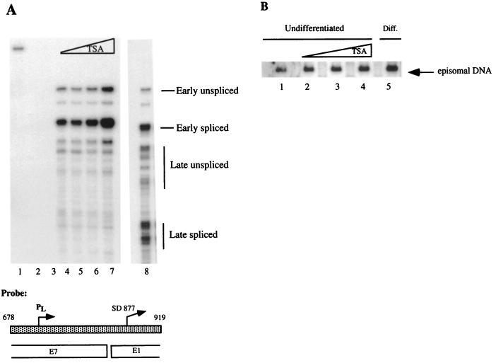
The life cycle of human papillomaviruses (HPVs) is tightly linked to the differentiation status of the host cell. While early genes are expressed during the initial stages of viral infection, late gene expression occurs in the suprabasal layers of the cervical epithelium. Late genes encode E1-E4, a cytosolic protein, and capsid proteins L1 and L2. We have mapped over 30 initiation sites for late transcripts and show that the transcripts initiate in a 200-nucleotide region within the E7 open reading frame. The mechanisms regulating the activation of late gene expression, however, are not yet understood. DNase I hypersensitivity analysis of HPV-31 chromatin in cell lines that maintain viral genomes extrachromosomally indicates that a major shift in nuclease digestion occurs upon differentiation. In undifferentiated cells, hypersensitive regions exist in the upstream regulatory region proximal to the E6 open reading frame. Upon differentiation, a region between nucleotides 659 and 811 in the E7 open reading frame becomes accessible to DNase I. These results indicate that the late transcript initiation region becomes accessible to transcription factor binding upon differentiation. Several complexes mediate chromatin rearrangement, and we tested whether histone acetylation was sufficient for late transcript activation. Treatment with the histone deacetylase inhibitor trichostatin A was found to be insufficient to activate late gene expression in undifferentiated cells. However, it did activate expression of early transcripts. These results suggest that chromatin remodeling around the late promoter occurs upon epithelial differentiation and that mechanisms in addition to histone deacetylation contribute to activation of late gene expression.


Figures

FIG. 1
FIG. 1
(A) Linear representation of the HPV-31 genome. Open reading frames are indicated underneath the map. PE refers to the early promoter (p97); PL refers to the late promoter. PolyA indicates polyadenylation sites. (B) Cartoon depicting late HPV-31 transcripts. PL designates the late promoter. Splice sites are indicated by the numbers below each transcript. Open reading frames are specified to the right of each message.
FIG. 2
FIG. 2
RNase protection assays. Total RNA was isolated from monolayer or differentiated cells using the Trizol reagent (Invitrogen, Carlsbad, Calif.) per the manufacturer's instructions. Antisense riboprobes labeled with 32P were synthesized using the Riboprobe transcription system from Promega. Probes were purified following gel electrophoresis in 6% acrylamide. RNase protection assay was performed as described by Stubenrauch et al. (50), with the following exceptions: 15 μg of RNA was used for each hybridization to 1.5 × 10⁵ cpm of probe, and the RNA was digested with 12-μg/ml RNase A and 60-U/ml RNase T1 (Roche Molecular Biochemicals, Indianapolis, Ind.). Labeled 100-bp ladder (Invitrogen) and a DNA sequencing ladder were run with samples as size markers. Lanes contain hybridization samples as follows: 1, undigested probe; 2, no RNA; 3, yeast tRNA; 4, RNA from monolayer CIN 612 cells; 5, RNA from CIN 612 cells induced to differentiate in methylcellulose for 24 h. (A) The pRP-p742 probe, which detects both early and late messages, has been described previously (50). The probe is illustrated by a stippled rectangle below the autoradiograph. Corresponding open reading frames are specified under the probe. PL designates the late promoter, while SD877 represents a splice donor site at nucleotide 877. Early and late transcripts are marked to the right of the bands. (B to D) RNase protection assay for mapping of late transcripts. All probes are specified below each autoradiograph. The probe used in panel B spans nucleotides 600 to 850; the probe in panel C spans nucleotides 600 to 756; the probe in panel D includes nucleotides 750 to 850. The numbers to the right of each band indicate the nucleotide location of each initiation site. The slowest-migrating band in lanes 4 and 5 in each panel corresponds to the fully protected probe.
FIG. 3
FIG. 3
Partial linear map of HPV-31 with corresponding open reading frames at the bottom. The portion of the E7 ORF from nucleotides 600 to 850 has been magnified to mark with arrows the initiation sites that were identified in this study. Initiation sites that show a high frequency of usage are underlined. Transcripts that are constitutively expressed are indicated by asterisks. Sequences in dashed boxes are initiator elements, and the CCAAT box is indicated by a dashed oval.
FIG. 4
FIG. 4
DNase I hypersensitivity analysis of HPV-31. (A) Schematic representation of the parental fragment, probe, and corresponding regions in HPV-31. P97, early promoter. URR and open reading frames are indicated by boxes. Restriction sites are indicated by italics: BlpI; BanII; A, AlwNI; S, SpeI; H, HpaI. (B) Autoradiograph corresponding to a DNase I hypersensitivity assay. Lanes 1 to 5, nuclei from undifferentiated CIN 612 cells. Lanes 6 to 10, nuclei from CIN 612 cells that were induced to differentiate in semisolid medium for 24 h. Nuclei were treated with a titration of DNase I corresponding to 10, 5, 2.5, 1.25, and 0 U/μl. Genomic DNA was digested with BlpI and AlwNI restriction endonucleases to yield a 2.7-kb parental fragment, and areas of hypersensitivity were observed by Southern blot analysis with a probe corresponding to the BanII-AlwNI fragment of HPV-31. Subgenomic marker fragments used for mapping areas of nuclease digestion are indicated at the left. Regions of the HPV-31 genome that show hypersensitivity are indicated to the right of the autoradiograph.
FIG. 5
FIG. 5
(A) RNase protection assay. Probe pRP-p742 was used to examine whether histone hyperacetylation induced late gene expression after treatment of undifferentiated CIN 612 cells with TSA (Sigma-Aldrich, St. Louis, Mo.). TSA was dissolved in ethanol and diluted directly into culture media to a final concentration of 100, 300, or 900 nM. The same volume of ethanol was added to the untreated samples. Monolayer cells were treated for 24 h and used directly to isolate RNA or induced to differentiate in semisolid medium for 24 h. Lane 1, undigested probe; lane 2, no RNA; lane 3, yeast tRNA; lane 4, untreated monolayer CIN 612 cells; lanes 5 to 7, undifferentiated CIN 612 cells treated with TSA at 100 nM (lane 5), 300 nM (lane 6), or 900 nM (lane 7). Lane 8 shows the pattern of late gene expression induced following suspension in methylcellulose for 24 h in the absence of TSA. Transcripts are labeled to the right of bands. The low levels of late transcript initiation in undifferentiated cells are due to a small percentage of cells that differentiate spontaneously (43). (B) Southern blot analysis for episome copy number after treatment with TSA. The Southern blot assay was performed as previously described (24). Briefly, 10 μg of sheared genomic DNA was digested with BglII (New England Biolabs), an enzyme that does not cut the HPV-31 genome, or with HpaI (New England Biolabs), a single cutter in HPV-31. The digests were separated in 0.8% agarose, transferred to a ZetaProbe GT membrane (Bio-Rad), and hybridized to an HPV-31 genomic probe at 42°C. Lanes: 1, untreated monolayer CIN 612 cells; 2 to 4, monolayer CIN 612 cells treated with 100, 300, and 900 nM TSA, respectively; 5, untreated CIN 612 cells that were induced to differentiate in semisolid medium for 24 h. All bands were quantified by PhosphorImager analysis.

References

    1. Ait-Si-Ali S, Polesskaya A, Filleur S, Ferreira R, Duquet A, Robin P, Vervish A, Trouche D, Cabon F, Harel-Bellan A. CBP/p300 histone acetyl-transferase activity is important for the G1/S transition. Oncogene. 2000;19:2430–2437. - PubMed
    1. Alexiadis V, Varga-Weisz P D, Bonte E, Becker P B, Gruss C. In vitro chromatin remodelling by chromatin accessibility complex (CHRAC) at the SV40 origin of DNA replication. EMBO J. 1998;17:3428–3438. - PMC - PubMed
    1. Baker C C, Howley P M. Differential promoter utilization by the bovine papillomavirus in transformed cells and productively infected wart tissues. EMBO J. 1987;6:1027–1035. - PMC - PubMed
    1. Baker C C, Phelps W C, Lindgren V, Braun M J, Gonda M A, Howley P M. Structural and transcriptional analysis of human papillomavirus type 16 sequences in cervical carcinoma cell lines. J Virol. 1987;61:962–971. - PMC - PubMed
    1. Becker P, Renkawitz R, Schutz G. Tissue-specific DNaseI hypersensitive sites in the 5′-flanking sequences of the tryptophan oxygenase and the tyrosine aminotransferase genes. EMBO J. 1984;3:2015–2020. - PMC - PubMed

Publication types

LinkOut - more resources