. 2001 Sep 17;194(6):769-79.
doi: 10.1084/jem.194.6.769.

Dendritic cells induce peripheral T cell unresponsiveness under steady state conditions in vivo

Affiliations

Dendritic cells induce peripheral T cell unresponsiveness under steady state conditions in vivo

D Hawiger et al. J Exp Med. .

Abstract

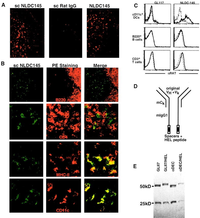
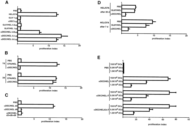
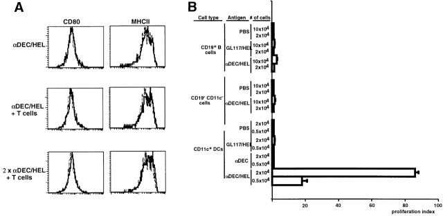
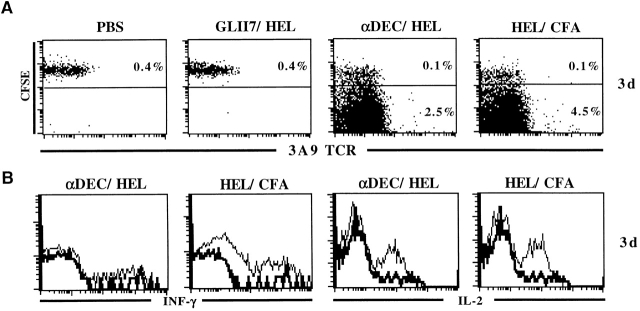
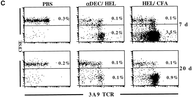
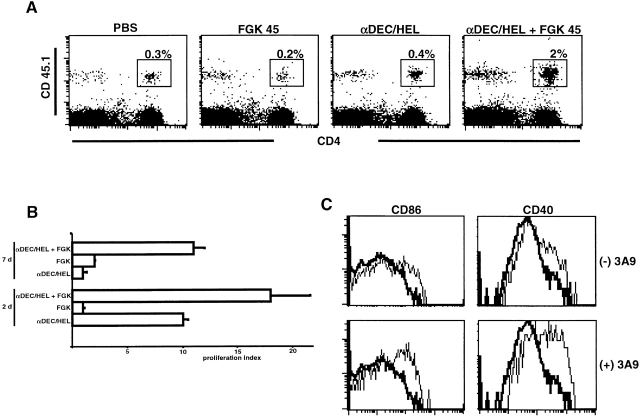
Dendritic cells (DCs) have the capacity to initiate immune responses, but it has been postulated that they may also be involved in inducing peripheral tolerance. To examine the function of DCs in the steady state we devised an antigen delivery system targeting these specialized antigen presenting cells in vivo using a monoclonal antibody to a DC-restricted endocytic receptor, DEC-205. Our experiments show that this route of antigen delivery to DCs is several orders of magnitude more efficient than free peptide in complete Freund's adjuvant (CFA) in inducing T cell activation and cell division. However, T cells activated by antigen delivered to DCs are not polarized to produce T helper type 1 cytokine interferon gamma and the activation response is not sustained. Within 7 d the number of antigen-specific T cells is severely reduced, and the residual T cells become unresponsive to systemic challenge with antigen in CFA. Coinjection of the DC-targeted antigen and anti-CD40 agonistic antibody changes the outcome from tolerance to prolonged T cell activation and immunity. We conclude that in the absence of additional stimuli DCs induce transient antigen-specific T cell activation followed by T cell deletion and unresponsiveness.


Figures

Figure 1
Figure 1
NLDC-145 targets DCs in vivo. (A) Biotinylated NLDC-145 (scNLDC145, left) or rat IgG (scRatIgG, middle) was injected into the hind footpads (50 μg/footpad) and inguinal LNs harvested 24 h later. Sections were stained with Streptavidin Cy3. Control sections from uninjected mice were stained using biotinylated NLDC145 and streptavidin Cy3 (NLDC145, right). (B) Two-color immunofluorescense. Mice were injected with biotinylated NLDC145 as in panel A. Sections were stained with streptavidin FITC (green) and PE-labeled antibodies (red) to B220 as indicated. Specimens were analyzed by deconvolution microscopy. Double labeling is indicated by the yellow color. (C) FACS® analysis of lymphoid cells 14 h after injection with NLDC145 and control GL117 antibody. Histograms show staining with anti–rat IgG on gated populations of CD11c+ DCs, B220+ B cells, and CD3+ T cells. (D) Diagrammatic representation of hybrid antibodies. (E) Hybrid antibodies. GL117, GL117/HEL, αDEC, and αDEC/HEL antibodies analyzed by PAGE under reducing conditions, molecular weights in kD are indicated.
Figure 3
Figure 3
In vivo activation of CD4+ T cells by αDEC/HEL. In all experiments, 3A9 T cells were transferred into B10.BR mice, and the recipients were injected subcutaneously in the footpads with antibodies in PBS or 100 μg of HEL peptide in CFA 24 h after T cell transfer as indicated. T cell proliferation was measured by [3H]thymidine incorporation and is expressed as a proliferation index relative to PBS controls. (A) T cells are efficiently activated by antigen delivered by αDEC/HEL. 48 h after challenge with antigen, CD4 T cells were isolated from peripheral LNs and cultured in vitro with irradiated B10.BR CD11c+ cells in the presence or absence of HEL peptide. (B) CD4+ T cells are only transiently activated by antigen (αDEC/HEL 0.2 μg) delivered to DCs in vivo. CD4+ cells were purified from peripheral LNs 2 or 7 d after challenge with antigen and cultured with irradiated CD11c+ cells in the presence or absence of HEL peptide. (C) Failure to induce persistent T cell activation with multiple injections of αDEC/HEL. 3A9 cells were transferred into B10.BR mice and recipients were injected with αDEC/HEL (0.2 μg/mouse) once (on day 9 or 2 before analysis) or multiple times (days 9, 6, and 2 before analysis). Assay for T cell activation was as above. (D) T cells initially activated by αDEC/HEL show diminished response to rechallenge with HEL peptide in CFA. Recipients were initially injected with either αDEC/HEL (0.2 μg), GL117/HEL(0.2 μg), or PBS and rechallenged 7 or 20 d later with 100 μg of HEL peptide in CFA or with PBS. CD4+ cells were purified from peripheral LNs (or spleens, not shown) 2 d after the rechallenge and cultured with irradiated CD11c+ cells in the presence or absence of HEL peptide. Assay for T cell activation was as above. (E) Antigen loading of DCs with αDEC/HEL. B10.BR mice with or without transferred 3A9 T cells, were injected subcutaneously with 0.2 μg αDEC/HEL or PBS either at 8 d (αDEC/HEL) or at 1 and 8 d (αDEC/HELX2) after transfer. Antigen loading was measured 1 d after the last dose of αDEC/HEL by purifying CD11c+ DCs from peripheral LNs and culturing with purified 3A9 T cells. The results are means of triplicate cultures from one of three similar experiments.
Figure 2
Figure 2
DCs process and present antigen delivered by hybrid antibodies. (A) MHC II and CD80 expression on DCs is not altered by multiple injections of αDEC/HEL and 3A9 T cells. B10.BR mice transferred with 3A9 T cells and controls were injected subcutaneously in the footpads with 0.2 μg αDEC/HEL or PBS either at 8 d (αDEC/HEL) or at 1 and 8 d (αDEC/HELX2) after transfer (similar results were obtained by intravenous injection of chimeric antibodies, data not shown). 24 h after the last αDEC/HEL injection, DCs were purified from peripheral LNs and analyzed by flow cytometry for expression of CD80 and MHC II. Dotted lines in histograms indicate PBS control. (B) αDEC/HEL delivers HEL peptide to DCs in vivo. B10.BR mice were injected subcutaneously into footpads with 0.3 μg of αDEC/HEL or GL117/HEL or αDEC or PBS as indicated. CD11c+, CD19+, and CD11cCD19 cells were isolated from draining LNs 24 h after antibody injection and assayed for antigen processing and presentation to purified 3A9 T cells in vitro. T cell proliferation was measured by [3H]thymidine incorporation and is expressed as a proliferation index relative to PBS controls. The results are means of triplicate cultures from one of four similar experiments.
Figure 4
Figure 4
CD4+ T cells divide in response to antigen presented by DCs in vivo, produce IL-2 but not IFN-γ, and are then rapidly deleted. (A) CFSE labeled CD45.1+ 3A9 T cells were transferred into B10.BR and 24 h later, the recipients were injected subcutaneously in the footpads with αDEC/HEL (0.2 μg), GL117/HEL (0.2 μg), HEL peptide in CFA, or PBS. CD4+ T cells were purified by negative selection from regional LNs 3 d after challenge with antigen and analyzed by flow cytometry. The plots show staining with 1G12 anti-3A9 and CFSE intensity on gated populations of CD4+CD45.1+ cells. The numbers indicate the percentage of CFSE high (undivided) and CFSE low (divided) CD4+ T cells. The results are from one of two similar experiments. (B) T cells produce IL-2 but not IFN-γ in response to antigens presented on DCs under physiological conditions. 3A9 cells were transferred into B10.BR mice and 24 h later the recipients were injected subcutaneously in the footpads with αDEC/HEL (0.2 μg), GL117/HEL (0.2 μg), HEL peptide in CFA. CD4+. Histograms show staining with anti–IL-2 and anti–IFN-γ on gated populations of 3A9+CD4+ cells. The thick lines indicate PBS control. (C) Same as in panel A but analysis performed 7 or 20 d after antigen administration.
Figure 4
Figure 4
CD4+ T cells divide in response to antigen presented by DCs in vivo, produce IL-2 but not IFN-γ, and are then rapidly deleted. (A) CFSE labeled CD45.1+ 3A9 T cells were transferred into B10.BR and 24 h later, the recipients were injected subcutaneously in the footpads with αDEC/HEL (0.2 μg), GL117/HEL (0.2 μg), HEL peptide in CFA, or PBS. CD4+ T cells were purified by negative selection from regional LNs 3 d after challenge with antigen and analyzed by flow cytometry. The plots show staining with 1G12 anti-3A9 and CFSE intensity on gated populations of CD4+CD45.1+ cells. The numbers indicate the percentage of CFSE high (undivided) and CFSE low (divided) CD4+ T cells. The results are from one of two similar experiments. (B) T cells produce IL-2 but not IFN-γ in response to antigens presented on DCs under physiological conditions. 3A9 cells were transferred into B10.BR mice and 24 h later the recipients were injected subcutaneously in the footpads with αDEC/HEL (0.2 μg), GL117/HEL (0.2 μg), HEL peptide in CFA. CD4+. Histograms show staining with anti–IL-2 and anti–IFN-γ on gated populations of 3A9+CD4+ cells. The thick lines indicate PBS control. (C) Same as in panel A but analysis performed 7 or 20 d after antigen administration.
Figure 5
Figure 5
CD40 ligation prolongs T cell activation in response to antigens delivered to DCs and induces upregulation of costimulatory molecules on DCs. (A) CD40 ligation induces persistent expansion of 3A9 cells in response to antigens delivered to DCs. CD45.1+ 3A9 T cells were transferred into B10.BR mice and 24 h later the recipients were injected subcutaneously in the footpads with 0.2 μg of αDEC/HEL alone or 90 μg of FGK45 or both or PBS. CD4+ T cells were purified by negative selection from regional LNs 7 d after challenge with antigen and analyzed by flow cytometry using antibodies specific for CD45.1 and CD4. The numbers indicate the percentages of CD4+CD45.1+ cells in LNs. (B) CD40 ligation prolongs T cell activation. 3A9 T cells were transferred into B10.BR mice and 24 h later, recipients were injected subcutaneously in the footpads with 0.2 μg of αDEC/HEL alone or 90 μg of FGK45 or both or PBS. After 2 or 7 d, CD4 T cells were isolated from the draining LNs and cultured in vitro with irradiated B10.BR CD11c+ cells in presence or absence of HEL peptide. T cell proliferation was measured by [3H]thymidine incorporation. The results represent triplicate cultures from two independent experiments. (C) CD40 ligation induces costimulatory molecules on DCs. B10.BR mice with or without 3A9 cell transfer were injected with 90 μg FGK45 plus 0.2 μg αDEC/HEL or αDEC/HEL or PBS. 3 d later DCs were isolated as in Fig. 2 and analyzed by flow cytometry using antibodies specific for CD11c, B220, CD86, and CD40. Histograms show staining with anti-CD40 and anti-CD86 on gated populations of DCs. Thick lines indicate control with PBS, which was same as αDEC/HEL alone.

References

    1. Banchereau J., Steinman R.M. Dendritic cells and the control of immunity. Nature. 1998;392:245–252. - PubMed
    1. Thery C., Amigorena S. The cell biology of antigen presentation in dendritic cells. Curr. Opin. Immunol. 2001;13:45–51. - PubMed
    1. Inaba K., Steinman R.M., Van Voorhis W.C., Muramatsu S. Dendritic cells are critical accessory cells for thymus-dependent antibody responses in mouse and in man. Proc. Natl. Acad. Sci. USA. 1983;80:6041–6045. - PMC - PubMed
    1. Steinman R.M., Gutchinov B., Witmer M.D., Nussenzweig M.C. Dendritic cells are the principal stimulators of the primary mixed leukocyte reaction in mice. J. Exp. Med. 1983;157:613–627. - PMC - PubMed
    1. Dhodapkar M.V., Steinman R.M., Sapp M., Desai H., Fossella C., Krasovsky J., Donahoe S.M., Dunbar P.R., Cerundolo V., Nixon D.F., Bhardwaj N. Rapid generation of broad T-cell immunity in humans after a single injection of mature dendritic cells. J. Clin. Invest. 1999;104:173–180. - PMC - PubMed

Publication types