. 2001 Sep 17;194(6):781-95.
doi: 10.1084/jem.194.6.781.

C1q and mannose binding lectin engagement of cell surface calreticulin and CD91 initiates macropinocytosis and uptake of apoptotic cells

Affiliations

C1q and mannose binding lectin engagement of cell surface calreticulin and CD91 initiates macropinocytosis and uptake of apoptotic cells

C A Ogden et al. J Exp Med. .

Abstract

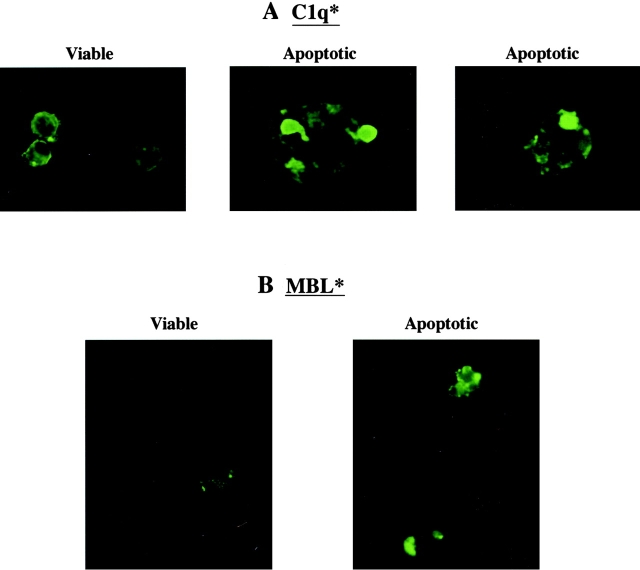
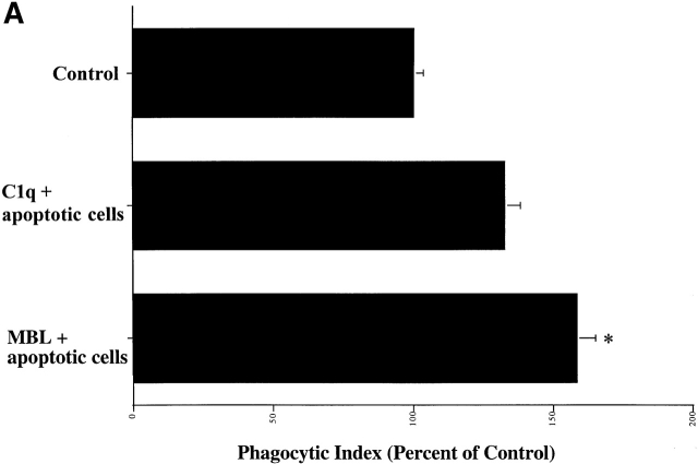
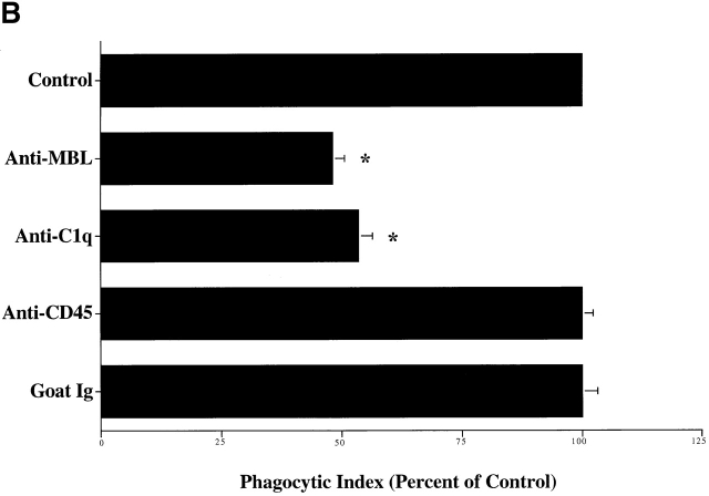
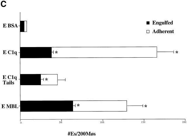
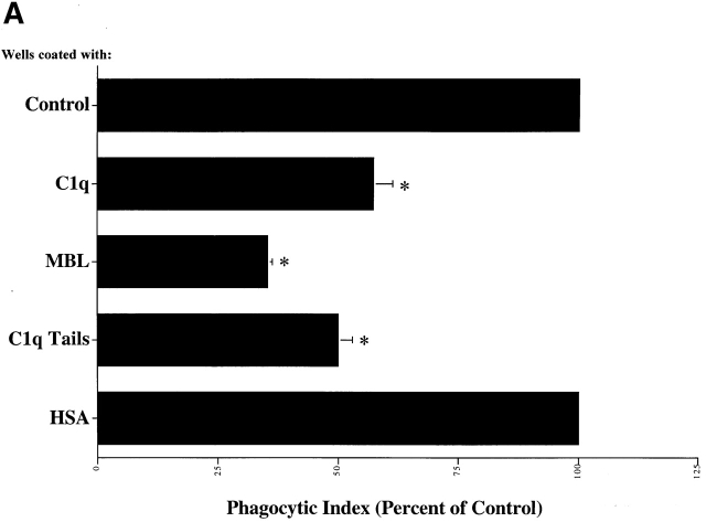
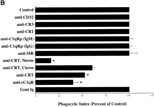
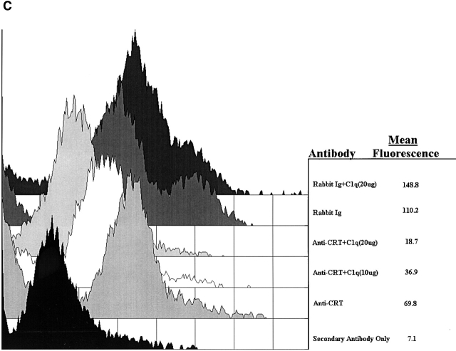
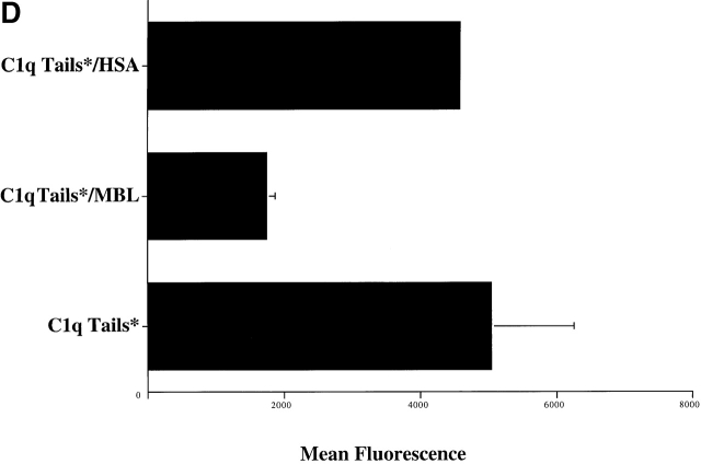
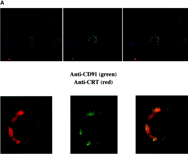
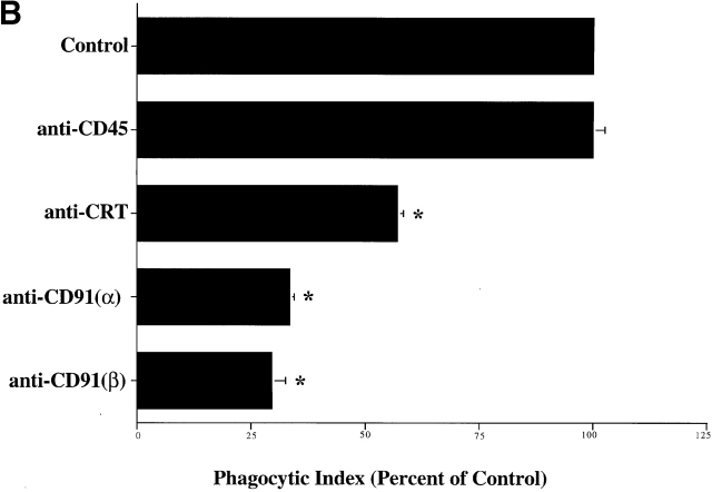
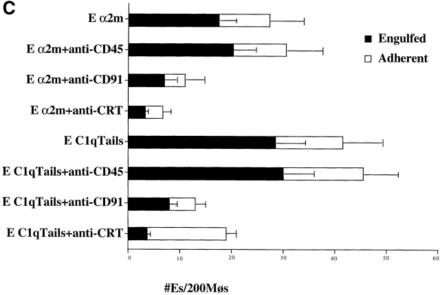
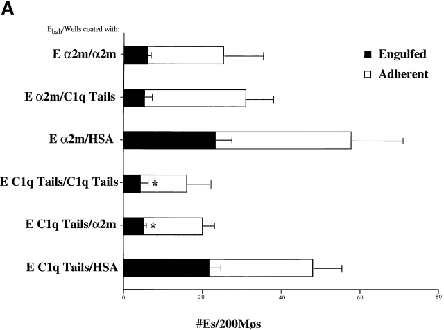
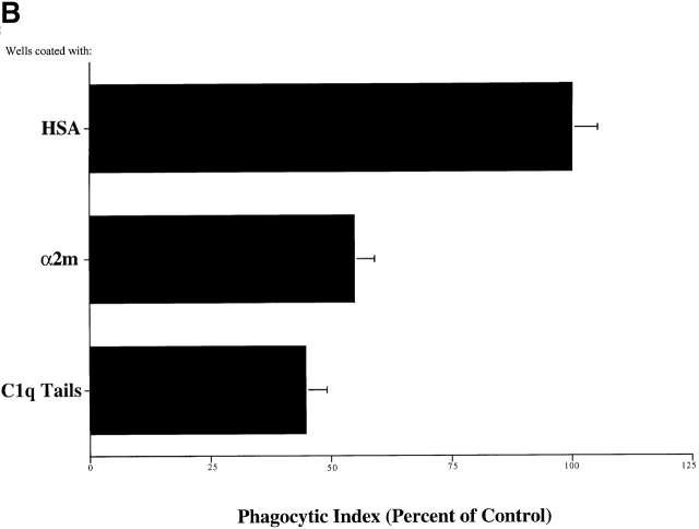
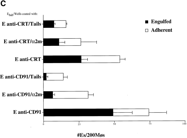
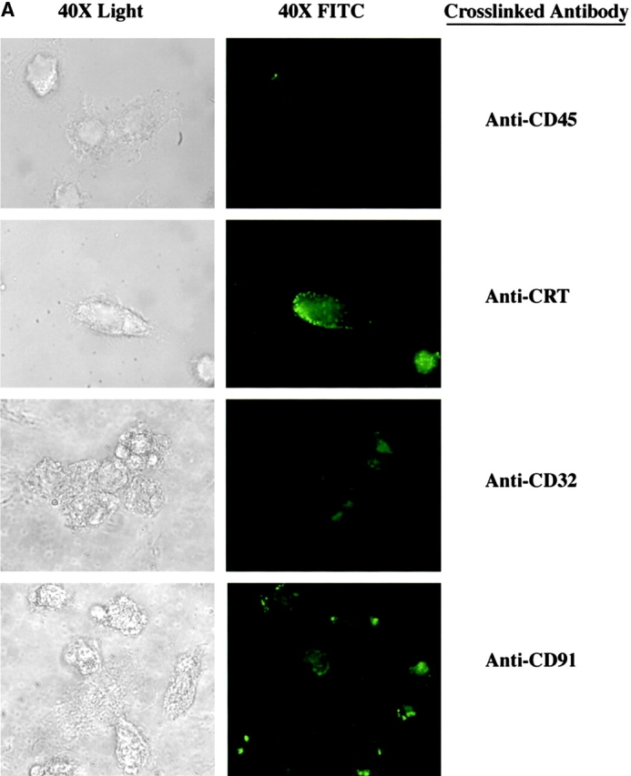
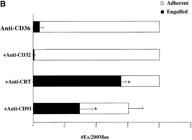
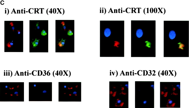
Removal of apoptotic cells is essential for maintenance of tissue homeostasis, organogenesis, remodeling, development, and maintenance of the immune system, protection against neoplasia, and resolution of inflammation. The mechanisms of this removal involve recognition of the apoptotic cell surface and initiation of phagocytic uptake into a variety of cell types. Here we provide evidence that C1q and mannose binding lectin (MBL), a member of the collectin family of proteins, bind to apoptotic cells and stimulate ingestion of these by ligation on the phagocyte surface of the multifunctional protein, calreticulin (also known as the cC1qR), which in turn is bound to the endocytic receptor protein CD91, also known as the alpha-2-macroglobulin receptor. Use of these proteins provides another example of apoptotic cell clearance mediated by pattern recognition molecules of the innate immune system. Ingestion of the apoptotic cells through calreticulin/CD91 stimulation is further shown to involve the process of macropinocytosis, implicated as a primitive and relatively nonselective uptake mechanism for C1q- and MBL-enhanced engulfment of whole, intact apoptotic cells, as well as cell debris and foreign organisms to which these molecules may bind.


Figures

Figure 1
Figure 1
C1q and MBL bind to the surface of apoptotic cells. Both Jurkat T cells and human neutrophils (not shown) bound C1q in their fresh and apoptotic states (A). C1q exhibited diffuse surface binding to the viable cells, while it bound in a localized pattern to apparent blebs on the surface of apoptotic cells. MBL showed little or no binding to fresh cells and robust, localized binding to the surface of apoptotic cells (B).
Figure 2
Figure 2
C1q and MBL facilitate particle clearance. (A) Pretreatment of apoptotic cells with C1q or MBL: apoptotic cells (5 × 10⁶) were incubated with 50 μg of purified C1q or MBL. These cells were tumbled for 30 min at 37°C and washed with HBSS before addition to macrophages for a phagocytosis assay (as described in Materials and Methods). n = 4 ± SEM (control mean phagocytic index = 44.2 ± 6.6, P < 0.05). (B) Antibody against human C1q and against human MBL inhibits uptake of apoptotic cells into HMDMs. HMDMs were treated with 10 μg antibody (polyclonal goat anti–human C1q, goat control antibody, mouse monoclonal anti-CD45 antibody as control, and monoclonal mouse anti–human MBL, shown) for 30 min at 37°C. Media was then aspirated, and apoptotic cells were added for 1 h for a phagocytosis assay. n = 4 ± SEM (control mean phagocytic index = 37.0 ± 6.1, *P < 0.005). (C) C1q, C1q Tails, or MBL Ebab are ingested by HMDMs. Single ligand coated particles coated with control protein, MBL, C1q, or the collagenous tail fragment of C1q were fed to HMDMs for 20 min at 37°C. The cells were then washed, fixed, and counted (as described in the Materials and Methods section). n = 3 ± SEM. P < 0.005 Engulfed; P < 0.003 Adherent.
Figure 2
Figure 2
C1q and MBL facilitate particle clearance. (A) Pretreatment of apoptotic cells with C1q or MBL: apoptotic cells (5 × 10⁶) were incubated with 50 μg of purified C1q or MBL. These cells were tumbled for 30 min at 37°C and washed with HBSS before addition to macrophages for a phagocytosis assay (as described in Materials and Methods). n = 4 ± SEM (control mean phagocytic index = 44.2 ± 6.6, P < 0.05). (B) Antibody against human C1q and against human MBL inhibits uptake of apoptotic cells into HMDMs. HMDMs were treated with 10 μg antibody (polyclonal goat anti–human C1q, goat control antibody, mouse monoclonal anti-CD45 antibody as control, and monoclonal mouse anti–human MBL, shown) for 30 min at 37°C. Media was then aspirated, and apoptotic cells were added for 1 h for a phagocytosis assay. n = 4 ± SEM (control mean phagocytic index = 37.0 ± 6.1, *P < 0.005). (C) C1q, C1q Tails, or MBL Ebab are ingested by HMDMs. Single ligand coated particles coated with control protein, MBL, C1q, or the collagenous tail fragment of C1q were fed to HMDMs for 20 min at 37°C. The cells were then washed, fixed, and counted (as described in the Materials and Methods section). n = 3 ± SEM. P < 0.005 Engulfed; P < 0.003 Adherent.
Figure 3
Figure 3
C1q and MBL facilitate clearance of apoptotic cells via cell-surface CRT. (A) Modulation of membrane receptor(s) by surface-bound C1q, C1q tails, or MBL. Macrophages were plated onto wells coated with control protein (HSA), MBL, C1q, or C1q tails. Apoptotic cells were then added for a phagocytosis assay. n = 7 ± SEM (control mean phagocytic index = 38.5 ± 4.4, P < 0.01). (B) Anti-CRT antibodies inhibit uptake of apoptotic cells by HMDMs. Other anti-receptor antibodies had no effect. anti-Nterm, anti-CRT, NH2 terminus; anti-Cterm, anti-CRT, COOH terminus; anti-MR, anti-mannose receptor; anti-CR3, anti-complement receptor 3; anti-CR1, anti-complement receptor 1. n = 4 ± SEM (control mean phagocytic index = 40.0 ± 4.3, P < 0.05). (C) Binding of anti-CRT antibody to the surface of HMDMs. Several polyclonal anti–human CRT antibodies were found to bind to the surface of HMDMs (see Materials and Methods) in a similar fashion. Binding of a rabbit anti–human polyclonal anti-CRT antibody was blocked by C1q. C1q did not inhibit binding of irrelevant antibody HMDM surfaces (inset). n = 3, representative experiment shown. (D) MBL and C1q bind to same receptor. C1q tails were FITC-conjugated (see Materials and Methods) and bound to HMDM surfaces. Unlabeled, whole MBL decreased this binding when incubated with the HMDMs along with the FITC-labeled tails, n = 3.
Figure 3
Figure 3
C1q and MBL facilitate clearance of apoptotic cells via cell-surface CRT. (A) Modulation of membrane receptor(s) by surface-bound C1q, C1q tails, or MBL. Macrophages were plated onto wells coated with control protein (HSA), MBL, C1q, or C1q tails. Apoptotic cells were then added for a phagocytosis assay. n = 7 ± SEM (control mean phagocytic index = 38.5 ± 4.4, P < 0.01). (B) Anti-CRT antibodies inhibit uptake of apoptotic cells by HMDMs. Other anti-receptor antibodies had no effect. anti-Nterm, anti-CRT, NH2 terminus; anti-Cterm, anti-CRT, COOH terminus; anti-MR, anti-mannose receptor; anti-CR3, anti-complement receptor 3; anti-CR1, anti-complement receptor 1. n = 4 ± SEM (control mean phagocytic index = 40.0 ± 4.3, P < 0.05). (C) Binding of anti-CRT antibody to the surface of HMDMs. Several polyclonal anti–human CRT antibodies were found to bind to the surface of HMDMs (see Materials and Methods) in a similar fashion. Binding of a rabbit anti–human polyclonal anti-CRT antibody was blocked by C1q. C1q did not inhibit binding of irrelevant antibody HMDM surfaces (inset). n = 3, representative experiment shown. (D) MBL and C1q bind to same receptor. C1q tails were FITC-conjugated (see Materials and Methods) and bound to HMDM surfaces. Unlabeled, whole MBL decreased this binding when incubated with the HMDMs along with the FITC-labeled tails, n = 3.
Figure 4
Figure 4
CD91 is involved in C1q- and MBL-mediated uptake via CRT. (A) CRT colocalizes with CD91 on the HMDM cell surface. HMDMs were stained with chicken anti–human CRT (anti–N-terminus, shown) and mouse anti–human CD91α and β (see Materials and Methods). Antibody staining was detected with FITC anti–mouse and Cy3 anti–chicken. n = 3, representative experiment shown. (B) Uptake of apoptotic cells is inhibited by antibody to CD91. HMDMs were preincubated with anti-CD91 antibody for 30 min at 37°C. Media was then aspirated and apoptotic cells were added for 1 h for a phagocytosis assay (see Materials and Methods). anti-CRT, chicken anti-CRT, NH2 terminus; anti-CD91α, anti-CD91, α chain; anti-CD91β, anti-CD91, β chain. n = 3 ± SEM (control mean phagocytic index = 20.3 ± 1.5, P < 0.002). (C) C1q tail Ebab and α2m Ebab are taken up into HMDMs; this uptake is inhibited by anti-CD91 and by anti-CRT. Ebab coated with C1q tails, α2m, or BSA (not shown) were fed to HMDMs for 20 min at 37°C (see Materials and Methods). n = 4. P < 0.1 Engulfed; P < 0.1 Adherent.
Figure 4
Figure 4
CD91 is involved in C1q- and MBL-mediated uptake via CRT. (A) CRT colocalizes with CD91 on the HMDM cell surface. HMDMs were stained with chicken anti–human CRT (anti–N-terminus, shown) and mouse anti–human CD91α and β (see Materials and Methods). Antibody staining was detected with FITC anti–mouse and Cy3 anti–chicken. n = 3, representative experiment shown. (B) Uptake of apoptotic cells is inhibited by antibody to CD91. HMDMs were preincubated with anti-CD91 antibody for 30 min at 37°C. Media was then aspirated and apoptotic cells were added for 1 h for a phagocytosis assay (see Materials and Methods). anti-CRT, chicken anti-CRT, NH2 terminus; anti-CD91α, anti-CD91, α chain; anti-CD91β, anti-CD91, β chain. n = 3 ± SEM (control mean phagocytic index = 20.3 ± 1.5, P < 0.002). (C) C1q tail Ebab and α2m Ebab are taken up into HMDMs; this uptake is inhibited by anti-CD91 and by anti-CRT. Ebab coated with C1q tails, α2m, or BSA (not shown) were fed to HMDMs for 20 min at 37°C (see Materials and Methods). n = 4. P < 0.1 Engulfed; P < 0.1 Adherent.
Figure 5
Figure 5
CD91 and CRT facilitate particle clearance. (A) α2m and C1q Tails modulate the uptake of tail Ebab and of α2m Ebab. HMDMs were plated onto wells coated with either HSA, α2m, or C1q tails (see Materials and Methods). These macrophages were then fed erythrocytes coated with C1q tails, α2m, or BSA (not shown) (α2m/HSA, for example, stands for α2m Ebab fed to macrophages plated onto wells coated with HSA). n = 3 ± SEM. P < 0.02 Engulfed. (B) α2m and C1q tails modulate the uptake of apoptotic cells. HMDMs were plated onto wells coated with HSA, α2m, or C1q tails. Apoptotic cells were fed to the macrophages for 1 h at 37°C for a phagocytosis assay. n = 3 ± SEM (control mean phagocytic index= 21.3 ± 5.3, P < 0.34). (C) α2m or C1q tails modulate the uptake of anti-CD91 or anti-CRT Ebab. HMDMs were plated onto wells coated with HSA, α2m, or C1q tails. The macrophages were then fed erythrocytes coated with BSA, anti-CD32 (neither shown), anti-CD91, or anti-CRT, NH2 terminus (here, anti-CRT). These Ebab were incubated with the HMDMs for 20 min. n = 3 ± SEM. P < 0.11 Engulfed; P < 0.06 Adherent.
Figure 5
Figure 5
CD91 and CRT facilitate particle clearance. (A) α2m and C1q Tails modulate the uptake of tail Ebab and of α2m Ebab. HMDMs were plated onto wells coated with either HSA, α2m, or C1q tails (see Materials and Methods). These macrophages were then fed erythrocytes coated with C1q tails, α2m, or BSA (not shown) (α2m/HSA, for example, stands for α2m Ebab fed to macrophages plated onto wells coated with HSA). n = 3 ± SEM. P < 0.02 Engulfed. (B) α2m and C1q tails modulate the uptake of apoptotic cells. HMDMs were plated onto wells coated with HSA, α2m, or C1q tails. Apoptotic cells were fed to the macrophages for 1 h at 37°C for a phagocytosis assay. n = 3 ± SEM (control mean phagocytic index= 21.3 ± 5.3, P < 0.34). (C) α2m or C1q tails modulate the uptake of anti-CD91 or anti-CRT Ebab. HMDMs were plated onto wells coated with HSA, α2m, or C1q tails. The macrophages were then fed erythrocytes coated with BSA, anti-CD32 (neither shown), anti-CD91, or anti-CRT, NH2 terminus (here, anti-CRT). These Ebab were incubated with the HMDMs for 20 min. n = 3 ± SEM. P < 0.11 Engulfed; P < 0.06 Adherent.
Figure 6
Figure 6
Uptake through CRT/CD91 occurs through a mechanism of macropinocytosis. (A) Stimulation of CRT and of CD91, but not of CD45 or CD32, initiate macropinocytosis. Macrophages were incubated in the presence of Lucifer Yellow. Anti-CD45 (above right) or anti-CRT, NH2 terminus (anti-CRT) (bottom right) were added to the macrophages (5 μg/ml) and allowed to interact for 15 min at room temperature. Cross-linking antibodies were then added (2.5 μg/ml), and the macrophages were incubated with Lucifer Yellow in the dark at 37°C for 5 min. Anti-CRT and anti-CD91 antibodies stimulated macropinocytosis and subsequent uptake of Lucifer Yellow dye, whereas anti-CD45 did not. (B) Bystander uptake of adherent particles by HMDMs occurs via CD91- and CRT-stimulated macropinocytosis. Chicken anti–human CRT (N-terminus) or mouse anti–human CD91 were used to stimulate HMDMs with anti-CD36–coated particles adherent to their surfaces. Cross-linking antibodies were then added to enhance the effect and phagocytosis was allowed to occur for 20 min. Each bar represents the total number of adherent cells plus engulfed cells. n = 5 ± SEM. P < 0.0001 Engulfed. (C) Engulfment of erythrocytes (E) coated with anti-CRT antibody involves formation of spacious phagosomes and concomitant uptake of Lucifer Yellow. RBCs labeled with anti-CRT, NH2 terminus (anti-CRT) (i and ii), anti-CD36 (iii), or anti-CD32 (iv) were stained with Texas red and added to HMDMs in the presence of Lucifer Yellow. Anti-CD36 RBCs (iii) were not taken up into the macrophages, anti-CD32 RBCs (iv) were ingested by macrophages, but with no accompanying uptake of Lucifer Yellow. In contrast, anti-CRT RBCs (i and ii) were taken up into HMDMs along with Lucifer Yellow, which colocalized in the same phagosome. HMDM nuclei stained with DAPI (blue).
Figure 6
Figure 6
Uptake through CRT/CD91 occurs through a mechanism of macropinocytosis. (A) Stimulation of CRT and of CD91, but not of CD45 or CD32, initiate macropinocytosis. Macrophages were incubated in the presence of Lucifer Yellow. Anti-CD45 (above right) or anti-CRT, NH2 terminus (anti-CRT) (bottom right) were added to the macrophages (5 μg/ml) and allowed to interact for 15 min at room temperature. Cross-linking antibodies were then added (2.5 μg/ml), and the macrophages were incubated with Lucifer Yellow in the dark at 37°C for 5 min. Anti-CRT and anti-CD91 antibodies stimulated macropinocytosis and subsequent uptake of Lucifer Yellow dye, whereas anti-CD45 did not. (B) Bystander uptake of adherent particles by HMDMs occurs via CD91- and CRT-stimulated macropinocytosis. Chicken anti–human CRT (N-terminus) or mouse anti–human CD91 were used to stimulate HMDMs with anti-CD36–coated particles adherent to their surfaces. Cross-linking antibodies were then added to enhance the effect and phagocytosis was allowed to occur for 20 min. Each bar represents the total number of adherent cells plus engulfed cells. n = 5 ± SEM. P < 0.0001 Engulfed. (C) Engulfment of erythrocytes (E) coated with anti-CRT antibody involves formation of spacious phagosomes and concomitant uptake of Lucifer Yellow. RBCs labeled with anti-CRT, NH2 terminus (anti-CRT) (i and ii), anti-CD36 (iii), or anti-CD32 (iv) were stained with Texas red and added to HMDMs in the presence of Lucifer Yellow. Anti-CD36 RBCs (iii) were not taken up into the macrophages, anti-CD32 RBCs (iv) were ingested by macrophages, but with no accompanying uptake of Lucifer Yellow. In contrast, anti-CRT RBCs (i and ii) were taken up into HMDMs along with Lucifer Yellow, which colocalized in the same phagosome. HMDM nuclei stained with DAPI (blue).

References

    1. Holmskov U., Malhotra R., Sim R.B., Jensenius J.C. Collectinscollagenous C-type lectins of the innate immune defense system. Immunol. Today. 1994;15:67–74. - PubMed
    1. Hansen S., Holmskov U. Structural aspects of collectins and receptors for collectins. Immunobiology. 1998;199:165–189. - PubMed
    1. Loos M., Martin H., Petry F. The biosynthesis of C1q, the collagen-like and Fc-recognizing molecule of the complement system. Behring Inst. Mitt. 1989;84:32–41. - PubMed
    1. Bowness P., Davies K.A., Norsworthy P.J., Athanassiou P., Taylor-Wiedeman J., Borysiewicz L.K., Meyer P.A., Walport M.J. Hereditary C1q deficiency and systemic lupus erythematosus. QJM. 1994;87:455–464. - PubMed
    1. Carroll M.C. The lupus paradox. Nat. Gen. 1998;19:3–4. - PubMed

Publication types

MeSH terms