. 2001 Sep 15;15(18):2408-20.
doi: 10.1101/gad.915901.

Identification of in vivo mRNA targets of GLD-1, a maxi-KH motif containing protein required for C. elegans germ cell development

Affiliations

Identification of in vivo mRNA targets of GLD-1, a maxi-KH motif containing protein required for C. elegans germ cell development

M H Lee et al. Genes Dev. .

Abstract

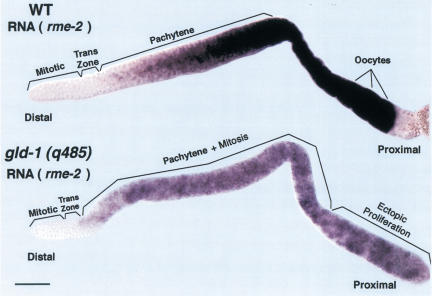
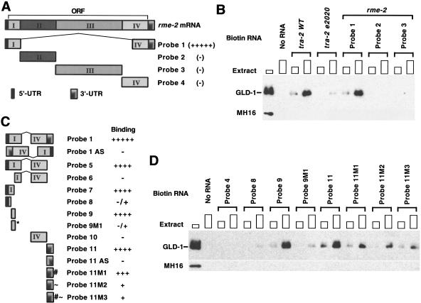
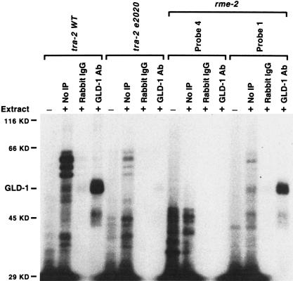
Caenorhabditis elegans GLD-1, a KH motif containing RNA-binding protein of the GSG/STAR subfamily, controls diverse aspects of germ line development, suggesting that it may have multiple mRNA targets. We used an immunoprecipitation/subtractive hybridization/cloning strategy to identify 15 mRNAs that are putative targets of GLD-1 binding and regulation. For one target, the rme-2 yolk receptor mRNA, GLD-1 acts as a translational repressor to spatially restrict RME-2 accumulation, and thus yolk uptake, to late-stage oocytes. We found that GLD-1 binds sequences in both 5' coding and the 3' untranslated region of rme-2 mRNA. Initial characterization of the other 14 targets shows that (1) they are coexpressed with GLD-1; (2) they can have mutant/RNA-mediated interference depletion phenotypes indicating functions in germ line development or as maternal products necessary for early embryogenesis; and (3) GLD-1 may coregulate mRNAs corresponding to functionally redundant subsets of genes within two gene families. Thus, a diverse set of genes have come under GLD-1-mediated regulation to achieve normal germ line development. Previous work identified tra-2 as a GLD-1 target for germ line sex determination. Comparisons of GLD-1-mediated translational control of rme-2 and tra-2 suggests that the mechanisms may differ for distinct target mRNA species.


Figures

Figure 1
Figure 1
Schematic representation of the adult hermaphrodite germ line and qualitative depiction of GLD-1 protein levels. Diagram of a single wild-type adult hermaphrodite gonad arm (upper panel) is drawn linearly instead of its normal reflexed shape for comparison purposes. Our qualitative assessment of GLD-1 protein levels (y-axis) in the corresponding regions of the germ line (x-axis) is shown in the lower panel. The gonad contains ∼1000 germ nuclei. In the distal region, nuclei are arranged primarily around the periphery of the gonadal tube. Each nucleus is partially enclosed by plasma membranes; although this is a syncytium, each nucleus and its surrounding cytoplasm and membranes is called a germ cell. See text for details.
Figure 2
Figure 2
(A) Procedure for identification of mRNA targets of GLD-1. (B) GLD-1/FLAG expression from animals rescued by complex arrays. Western blot of total extracts from wild type (+/+), gld-1 null heterozygote (q485/+), gld-1 null homozygote (q485), gld-1 null homozygote rescued with an untagged gld-1 genomic clone (q485;ozEx38), and gld-1 homozygote rescued with a FLAG-tagged gld-1 genomic clone (q485;ozEx40 and q485;ozEx41) probed with anti-GLD-1 (top panel), anti-FLAG (middle panel), or anti-β-tubulin antibodies (n357, Amersham) as a loading control. Each lane contains 100 adult hermaphrodites of the given genotype. In rescued lines, adult hermaphrodites that have at least one rescued gonad arm were picked. (C) Specific immunoprecipitation and elution of GLD-1/FLAG from the cytosol extract. Western blot of eluted proteins with FLAG peptide (E1–E5) after immunoprecipitation with nonimmune total mouse IgG (IgG IP) or with anti-FLAG antibody (FLAG IP) from the cytosol extract of q485; ozEx40 adult hermaphrodites. Flow through (F.T.) is the material that remained unbound after IgG IP or FLAG IP. The blot was probed with anti-GLD-1. Cytosol extract from wild-type adult hermaphrodites (WT) was loaded to show the untagged, endogenous GLD-1.
Figure 3
Figure 3
RME-2 yolk receptor accumulation is regulated by GLD-1. Gonad arms, dissected from a wild-type adult hermaphrodite (A,B) or from a gld-1 null adult hermaphrodite (C,D), stained with DAPI to visualize DNA (A,C as white), rat anti-GLD-1 (B as green), and rabbit anti-RME-2 (B,D as red). (B) GLD-1 and RME-2 accumulation is mutually exclusive. Composite shows an interior focal plane of an intact gonad. GLD-1 staining is the strongest in the transition zone and pachytene region. At the loop, as GLD-1 staining diminishes rapidly, RME-2 staining starts to appear. RME-2 staining becomes stronger and localized at the plasma membrane, as proximal oocytes become more fully cellularized and increase in volume. (D) RME-2 is misexpressed and localized at the plasma membrane of pachytene-stage germ cells in the distal region of the gld-1 null adult hermaphrodite germ line. At the proximal end, as germ cells proliferate ectopically, RME-2 staining is variable. Bar, 20 μm.
Figure 4
Figure 4
Accumulation of rme-2 mRNA in wild-type and gld-1 null adult hermaphrodite germ lines. A dissected wild-type adult hermaphrodite gonad (upper panel) showing rme-2 mRNA (purple staining) is first detected in early meiotic prophase, in the transition zone, and progressively increases to a high level in late-stage oocytes. A dissected gld-1 null adult hermaphrodite gonad (lower panel) showing rme-2 mRNA is first detected in early meiotic prophase and then increases to a higher level in pachytene that is maintained through the proximal tumorous region. The level of rme-2 RNA appears lower in gld-1 null than wild-type hermaphrodites, possibly because GLD-1 partially stabilizes the mRNA or transcription of rme-2 is reduced in tumorous germ cells. Bar, 20 μm.
Figure 5
Figure 5
GLD-1 binds to sequences at both the 5′ and the 3′ ends of rme-2 mRNA. (A,C) Schematic diagram of RNA probes. (B,D) Each biotin-labeled RNA or control sample (No RNA) was allowed to form a complex with increasing amounts of cytosol extract (∼50 μg or ∼150 μg of total protein) from wild-type adult hermaphrodites and the complex isolated with streptavidin magnetic beads. The isolated proteins were subjected to Western analysis with anti-GLD-1 antibody. The first lanes show total GLD-1 in cytosol extract (∼30 μg). The tra-2 WT 3′UTR and the tra-2(e2020) mutant 3′UTR (B) are control probe RNAs that should bind and not bind GLD-1, respectively. MH16 (anti-Paramyosin antibody) was used as a negative control. Probe 1 in A and C is identical; Probe 1 AS and Probe 11 AS are antisense versions. For the nucleotide positions of each rme-2 RNA fragment used as a probe for GLD-1 binding, see our web site: (http://www.genetics.wustl.edu/tslab/leesup.html).
Figure 6
Figure 6
GLD-1 directly contacts rme-2 RNA. 32P-labeled RNAs, tra-2 wild-type (WT) 3′UTR, tra-2(e2020) 3′UTR, or rme-2 Probe 4 or Probe 1 were incubated with (+) or without (−) cytosol extract (∼60 μg of protein) from wild-type adult hermaphrodites, cross-linked with UV, digested with RNase A, and immunoprecipitated with rabbit IgG (Sigma) or with anti-GLD-1 antibody (GLD-1 Ab). (No IP lanes) loaded with 10% of total material used in the experiment; (Rabbit IgG and GLD-1 Ab lanes) loaded with 100% of material after immunoprecipitation. Molecular masses and GLD-1 are indicated. Similar results were obtained with Probe 9 or Probe 11. We note that there are several polypeptides that bind the wild-type 3′UTR of tra-2 but are impaired in binding the 3′UTR of tra-2(e2020). They may represent RNA binding proteins that participate in translational repression through the 3′UTR or may regulate the reported localization of tra-2 mRNA (Graves et al. 1999).
Figure 7
Figure 7
RNA-binding activity of GLD-1 mutants correlates with their translational repression activity in vivo and their phenotype. Each biotin-labeled RNA was allowed to form a complex with increasing amounts of cytosol extracts (∼50 μg or ∼150 μg of proteins) from gld-1(q361); ozEx40 adult hermaphrodites (A) or from females and males of q126/q485 trans-heterozygote (B) and isolated with streptavidin magnetic beads. The isolated proteins were subjected to Western analysis with anti-GLD-1 antibody. The first lanes show total GLD-1(q361) (A) or GLD-1(q126) (B) in cytosol extract (∼30 μg). Since gld-1(q361); ozEx40 animals have GLD-1(q361) at normal levels but GLD-1/FLAG at less than one-twentieth of the normal level, GLD-1 found in A is essentially all the GLD-1(q361). We estimate that GLD-1(q126) forms a complex with the tra-2 3′UTR at one-fifth to one-tenth of the efficiency as wild-type GLD-1. Decreased binding of GLD-1(q126) to the tra-2 3′UTR, and thus presumably increased TRA-2 accumulation, may in part account for the feminized hermaphrodite germ line phenotype of gld-1(q126). However, because gld-1(q126) mutants also show feminization of the tra-2 null male germ line, GLD-1(q126) must have an additional tra-2 independent defect (Francis et al. 1995b).

References

    1. Austin J, Kimble J. glp-1 is required in the germ line for regulation of the decision between mitosis and meiosis in C. elegans. Cell. 1987;51:589–599. - PubMed
    1. Bowerman B, Draper B, Mello C, Priess J. The maternal gene skn-1 encodes a protein that is distributed unequally in early C. elegans embryos. Cell. 1993;74:443–452. - PubMed
    1. Chang L, Karin M. Mammalian MAP kinase signalling cascades. Nature. 2001;410:37–40. - PubMed
    1. Chen T, Damaj B, Herrerra C, Lasko P, Richard S. Self-association of the single-KH domain family members Sam68, GRP33, GLD-1 and Qk1: Role of the KH domain. Mol Cell Biol. 1997;17:5707–5718. - PMC - PubMed
    1. Church DL, Guan KL, Lambie EJ. Three genes of the MAP kinase cascade, mek-2, mpk-1/sur-1, and let-60 ras are required for meiotic cell cycle progression in Caenorhabditis elegans. Development. 1995;121:2525–2535. - PubMed

Publication types