Comparative Study
. 2001 Sep 25;98(20):11218-23.
doi: 10.1073/pnas.201220098. Epub 2001 Sep 18.

Structure of rat BCKD kinase: nucleotide-induced domain communication in a mitochondrial protein kinase

Affiliations
Comparative Study

Structure of rat BCKD kinase: nucleotide-induced domain communication in a mitochondrial protein kinase

M Machius et al. Proc Natl Acad Sci U S A. .

Abstract

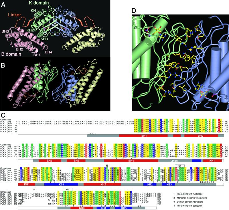
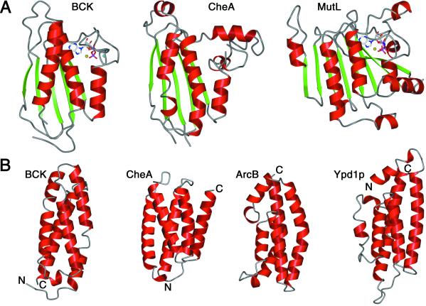
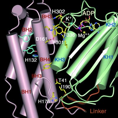
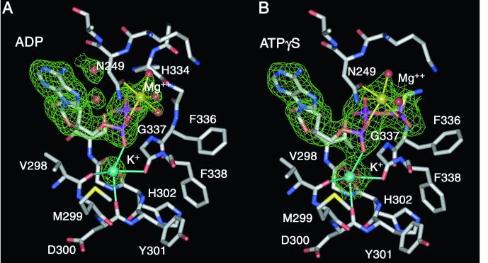
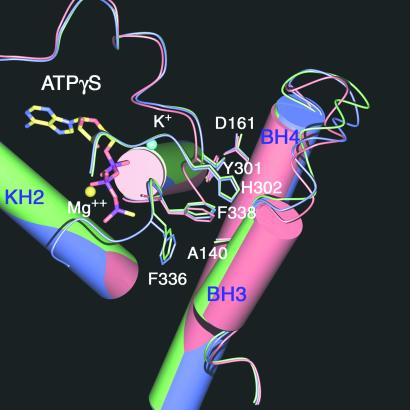
Mitochondrial protein kinases (mPKs) are molecular switches that down-regulate the oxidation of branched-chain alpha-ketoacids and pyruvate. Elevated levels of these metabolites are implicated in disease states such as insulin-resistant Type II diabetes, branched-chain ketoaciduria, and primary lactic acidosis. We report a three-dimensional structure of a member of the mPK family, rat branched-chain alpha-ketoacid dehydrogenase kinase (BCK). BCK features a characteristic nucleotide-binding domain and a four-helix bundle domain. These two domains are reminiscent of modules found in protein histidine kinases (PHKs), which are involved in two-component signal transduction systems. Unlike PHKs, BCK dimerizes through direct interaction of two opposing nucleotide-binding domains. Nucleotide binding to BCK is uniquely mediated by both potassium and magnesium. Binding of ATP induces disorder-order transitions in a loop region at the nucleotide-binding site. These structural changes lead to the formation of a quadruple aromatic stack in the interface between the nucleotide-binding domain and the four-helix bundle domain, where they induce a movement of the top portion of two helices. Phosphotransfer induces further ordering of the loop region, effectively trapping the reaction product ADP, which explains product inhibition in mPKs. The BCK structure is a prototype for all mPKs and will provide a framework for structure-assisted inhibitor design for this family of kinases.


Figures

Figure 1
Figure 1
The mammalian protein kinase BCK. (A) Overall view of the rat BCK dimer with the phosphotransfer reaction product ADP shown as ball-and-stick model. Strands in the K domain are not labeled. (B) Same as A, but rotated 90° around the horizontal line. All figures were made with bobscript (46) and povray (www.povray.org). (C) Sequence alignment of mitochondrial protein kinases. Similar residues are colored (yellow, hydrophobic; blue, basic; red, acidic; green, others). Residues observed in the rat BCK structure are depicted below the sequences (red, helices; blue, strands; gray, others); the conserved nucleotide-binding motifs are indicated above the consensus sequence. (D) Residues in the rat BCK dimer interface. Water molecules participate in dimer formation only at the periphery, but not in the core.
Figure 2
Figure 2
Comparison of BCK with structurally related kinases and ATPases. (A) Nucleotide-binding domain. BCK with ADP, CheA “empty” (PDB ID code 1B3Q), and MutL with ADPNP (PDB ID code 1B63). (B) B domain. BCK, CheA-HPt (PDB ID code 1i5n), ArcB (PDB ID code 1A0B), and Ypd1p (PDB ID code 1C02).
Figure 3
Figure 3
Residues in the interface between domains B (pink) and K (green) in the ADP-bound state of the rat BCK monomer (carbon atoms in yellow) and in the hydrophobic core of the B domain (carbon atoms in cyan).
Figure 4
Figure 4
Nucleotide-binding pocket of rat BCK with superimposed 2FoFc simulated annealing omit electron density maps (green, contoured at 1.5 σ) calculated for potassium (cyan) and magnesium (yellow), as well as bound ADP (A) and ATPγS (B). An anomalous difference Fourier electron density map (red, contoured at 5 σ) is shown for the ATPγS-bound BCK structure. The side chain of Phe-303 has been removed for clarity.
Figure 5
Figure 5
Superposition of the apo-structure of rat BCK (red) with its ADP-bound (blue) and ATPγS-bound form (green). Nucleotide (ADP or ATPγS) binding orders H302 and F336 (electron density not visible in the apo-form) in the B/K domain interface to form a quadruple aromatic stack between Y301, H302, F336, and F338. Structural differences in the BH3 and BH4 helices between the apo and the nucleotide-bound states are depicted.

References

    1. Chuang D T, Shih V E. In: The Metabolic and Molecular Basis of Inherited Disease. Scriver C R, Beaudet A L, Sly W S, Valle D, editors. New York: McGraw–Hill; 2001. pp. 1971–2006.
    1. Pettit F H, Yeaman S J, Reed L J. Proc Natl Acad Sci USA. 1978;75:4881–4885. - PMC - PubMed
    1. Chuang D T, Hu C W, Ku L S, Markovitz P J, Cox R P. J Biol Chem. 1985;260:13779–13786. - PubMed
    1. Paxton R, Harris R A. J Biol Chem. 1982;257:14433–14439. - PubMed
    1. Damuni Z, Merryfield M L, Humphreys J S, Reed L J. Proc Natl Acad Sci USA. 1984;81:4335–4338. - PMC - PubMed

Publication types

MeSH terms

Substances