. 2001 Sep;134(2):359-69.
doi: 10.1038/sj.bjp.0704251.

Bupivacaine effects on hKv1.5 channels are dependent on extracellular pH

Affiliations

Bupivacaine effects on hKv1.5 channels are dependent on extracellular pH

M Longobardo et al. Br J Pharmacol. 2001 Sep.

Abstract

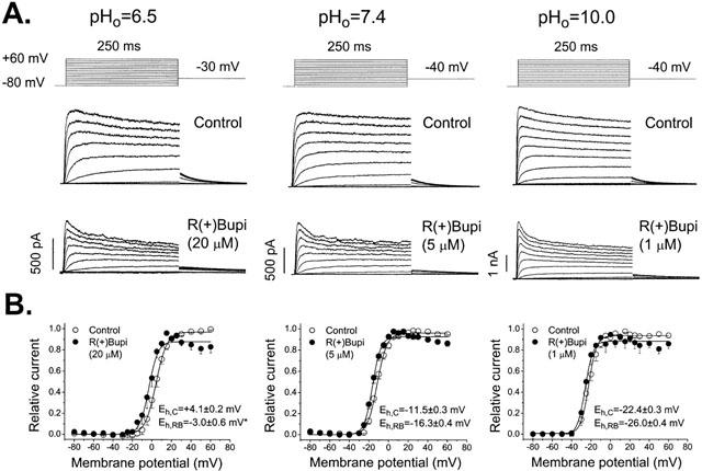
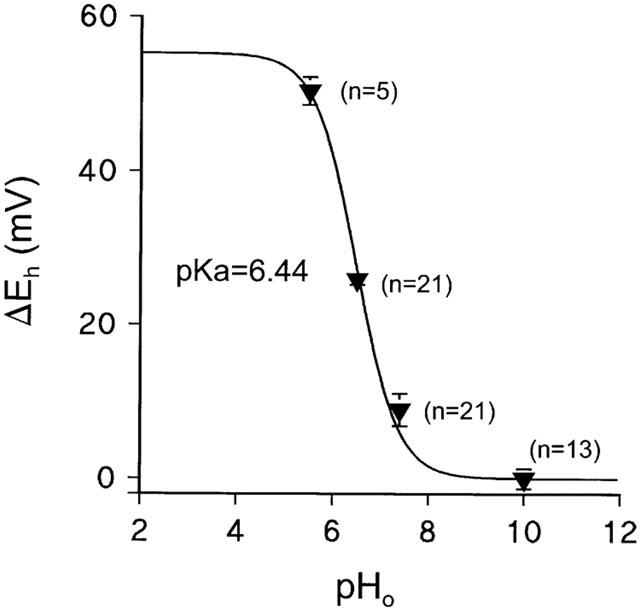
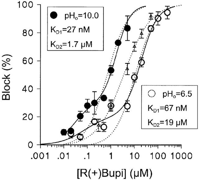
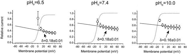
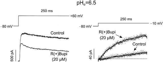
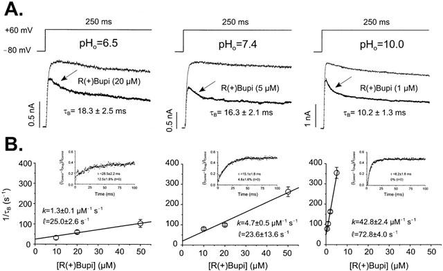
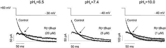
1. Bupivacaine-induced cardiotoxicity increases in hypoxic and acidotic conditions. We have analysed the effects of R(+)bupivacaine on hKv1.5 channels stably expressed in Ltk(-) cells using the whole-cell patch-clamp technique, at three different extracellular pH (pH(o)), 6.5, 7.4 and 10.0. 2. Acidification of the pH(o) from 7.4 to 6.5 decreased 4 fold the potency of R(+)bupivacaine to block hKv1.5 channels. At pH(o) 10.0, the potency of the drug increased approximately 2.5 fold. 3. Block induced by R(+)bupivacaine at pH(o) 6.5, 7.4 and 10.0, was voltage- and time-dependent in a manner consistent with an open state block of hKv1.5 channels. 4. At pH(o) 6.5, but not at pH(o) 7.4 or 10.0, R(+)bupivacaine increased by 95+/-3 % (n=6; P<0.05) the hKv1.5 current recorded at -10 mV, likely due to a drug-induced shift of the midpoint of activation (DeltaV=-8.5+/-1.4 mV; n=7). 5. R(+)bupivacaine development of block exhibited an 'instantaneous' component of block at the beginning of the depolarizing pulse, which averaged 12.5+/-1.8% (n=5) and 4.6+/-1.6% (n=6), at pH(o) 6.5 and 7.4, respectively, and that was not observed at pH(o) 10.0. 6. It is concluded that: (a) alkalinization of the pH(o) increases the potency of block of R(+)bupivacaine, and (b) at pH(o) 6.5, R(+)bupivacaine induces an 'agonist effect' of hKv1.5 current when recorded at negative membrane potentials.


Figures

Figure 1
Figure 1
Effects of R(+)bupivacaine on hKv1.5 channels at different pHo values. Original records obtained in the absence and in the presence of equipotent drug concentrations after applying depolarizing pulses from −80 to +60 mV in 10 mV steps of 250 ms duration. Tail currents were recorded upon repolarization to −40 mV (pHo 7.5 and 10.0) or −30 mV (pHo 6.5). (A) Shows original records obtained at pHo 6.5, 7.4 and 10.0 in the absence and in the presence of equipotent concentrations of R(+)bupivacaine. R(+)bupivacaine 20, 5 and 1 μM induced a time-dependent block which yields 62±4% (n=6), 61±2% (n=5) and 47±5% (n=5) at pHo 6.5, 7.4 and 10.0, respectively. (B) Shows the activation curves at pHo 6.5, 7.4 and 10.0 in the absence and in the presence of equipotent R(+)bupivacaine concentrations. The midpoint of activation (Eh) was shifted to more negative potentials as the pHo value was more alkaline. Data points represent the mean±s.e.mean of a 6 – 7 experiments. *P<0.01.
Figure 2
Figure 2
Effects of changes in extracellular pH (pHo) on hKv1.5 channels. Considering the equation ΔEh=A/(1+10(pH-pKa)); where A represents the maximum amplitude, and assuming that Eh saturates at pHo 10.0 (so ΔEh=0 at pHo=10.0), we calculated a pKa value of 6.44. This is in close agreement with the pKa value of protonation for an histidine (6.0) and suggests that the titration of a histidine present within the pore region of hKv1.5 channels could be responsible for the observed proton-induced shift towards more positive membrane potentials as the pHo is reduced.
Figure 3
Figure 3
Concentration-response curves for R(+)bupivacaine-induced block at all the pHo studied. In this plot we represented the R(+)bupivacaine induced block at each concentration and under each experimental conditions. At pHo 6.5 and 10.0 the concentration-response curves were better fit assuming a Hill equation of two components, i.e. biphasic concentration-response curves, from which two KD values were obtained (KD1 and KD2). Dotted lines represent the concentration-response curves when the fit assumed a Hill equation of one component. The concentration-response curve for R(+)bupivacaine at pHo 7.4 (Δ) was taken from (Valenzuela et al., 1995a) with permission.
Figure 4
Figure 4
Voltage-dependent effects of equipotent R(+)bupivacaine concentrations on hKv1.5 channels at different pHo values. Relationship between the relative current (IDrug/IControl) and membrane potential at pHo 6.5, 7.4 and 10.0. At the three pHo studied, block induced by R(+)bupivacaine steeply increased in the range of activation of the channel. At voltages positive to +10 mV (pHo 6.5), 0 mV (pHo 7.4) and −5 mV (pHo 10.0) block increased with a shallower voltage-dependence consistent with δ values of 0.18±0.01 (n=14), 0.18±0.01 (n=5) and 0.16±0.01 (n=7), respectively (P>0.05).
Figure 5
Figure 5
Effects of R(+)bupivacaine on hKv1.5 channels at pHo 6.5. Left panel shows original records obtained after depolarizing the cell from −80 to +60 mV. Right panel shows the drug effects after depolarizing the cell membrane to −10 mV. Note that, under these circumstances, the drug increased the amplitude of the hKv1.5 current by 95±3% (n=6; P<0.05), whereas after strong depolarizations (left panel), R(+)bupivacaine inhibited the hKv1.5 current.
Figure 6
Figure 6
Time-dependent block of hKv1.5 channels induced by R(+)bupivacaine at pHo 6.5, 7.5 and 10.0. (A) Shows original records obtained after applying a depolarizing pulse from −80 mV to +60 mV for 250 ms in duration in the absence and in the presence of R(+)bupivacaine (20, 5 and 1 μM) at the different pHo values. R(+)bupivacaine induced a fast initial decline of the current which was faster at higher drug concentrations and thus, it was considered as a good index of the time constant of block (τBlock). (B) The inverse of τBlock was plotted versus bupivacaine concentration. For a first-order blocking scheme, a linear relation is expected: τBlock−1=k×[D]+l (see Methods section). The solid line represents the linear fit, from which the apparent binding and unbinding rate constants were obtained. Inset: Representative traces of time course of the development of block of hKv1.5 after a depolarizing pulse to +60 mV from a holding potential of −80 mV at pHo 6.5, 7.4 and 10.0 induced by R(+)bupivacaine at concentrations of 10, 5 and 1 μM, respectively. The reduction of hKv1.5 current in the presence of the drug is expressed as a proportion of the control current at any given time after the beginning of the depolarizing pulse. In the presence of R(+)bupivacaine at all the pHo studied inhibition of the current increases exponentially during depolarization. Note that at pHo 6.5 and 7.4, R(+)bupivacaine (10 μM) induced an instantaneous block at t=0 ms.
Figure 7
Figure 7
Time-dependent block of hKv1.5 channels induced by R(+)bupivacaine at different pHo values observed during deactivation at −40 mV (at pHo 7.4 and 10.0) and −30 mV (at pHo 6.5). R(+)bupivacaine induced an initial rising phase due to the unblocking of R(+)bupivacaine from open channels and slowed the deactivation time course at all the pHo studied which resulted in a crossover phenomenon indicative of an open-channel block mechanism. The arrows indicate the ‘crossover' between the tail currents recorded in the presence of drug and those recorded under control conditions.

Similar articles

Cited by

References

    1. ANUMONWO J.M., HORTA J., DELMAR M., TAFFET S.M., JALIFE J. Proton and zinc effects on HERG currents. Biophys. J. 1999;77:282–298. - PMC - PubMed
    1. ARMSTRONG C.M. Interaction of tetraethylammonium ion derivatives with the potassium channels of giant axons. J. Gen. Physiol. 1971;58:413–437. - PMC - PubMed
    1. ÅBERG G. Toxicological and local anaesthetic effects of optically active isomers of two local anaesthetic compounds. Acta Pharmacol. Toxicol. 1972;31:273–286. - PubMed
    1. BETHELL H.W., VANDENBERG J.I., SMITH G.A., GRACE A.A. Changes in ventricular repolarization during acidosis and low- flow ischemia. Am. J. Physiol. 1998;275:H551–H561. - PubMed
    1. BUSCH A.E., HURST R.S., NORTH R.A., ADELMAN J.P., KAVANAUGH M.P. Current inactivation involves a histidine residue in the pore of the rat lymphocyte potassium channel RGK5. Biochem. Biophys. Res. Comm. 1991;179:1384–1390. - PubMed

Publication types