Comparative Study
. 2001 Sep 17;154(6):1125-33.
doi: 10.1083/jcb.200106011.

Eg5 is static in bipolar spindles relative to tubulin: evidence for a static spindle matrix

Affiliations
Comparative Study

Eg5 is static in bipolar spindles relative to tubulin: evidence for a static spindle matrix

T M Kapoor et al. J Cell Biol. .

Abstract

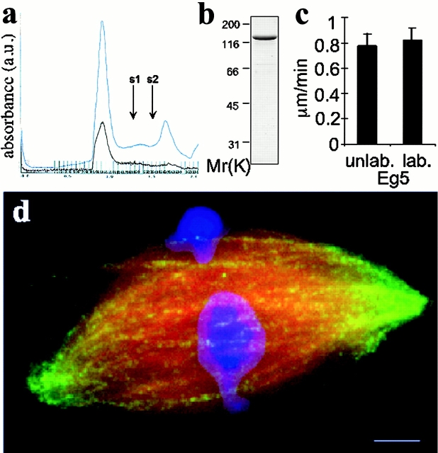
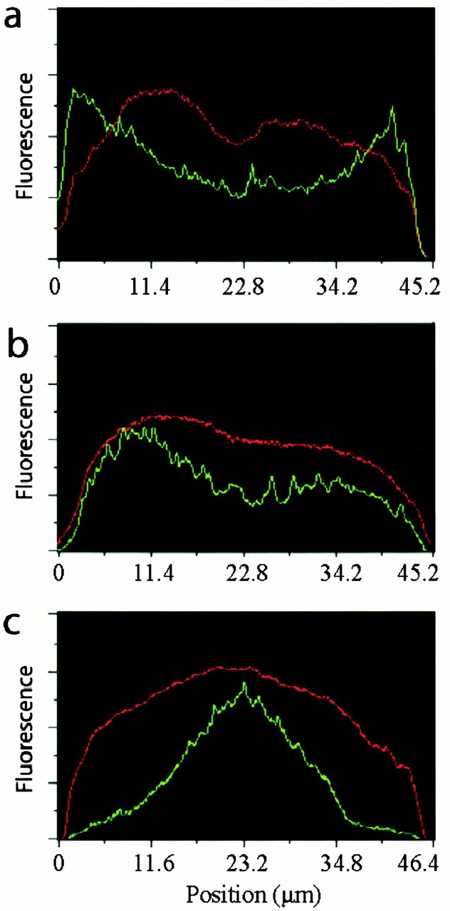
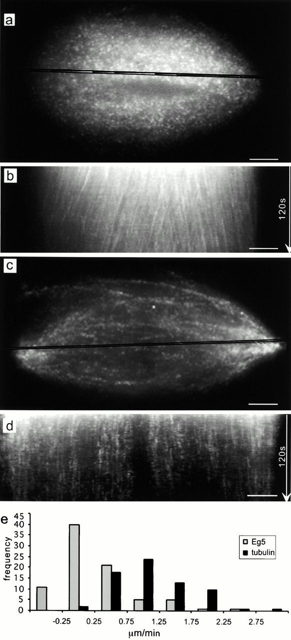
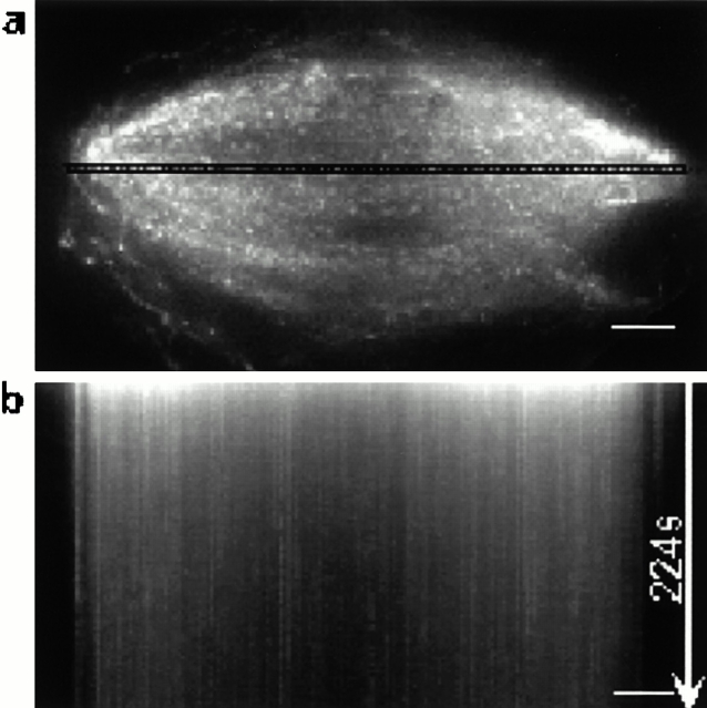
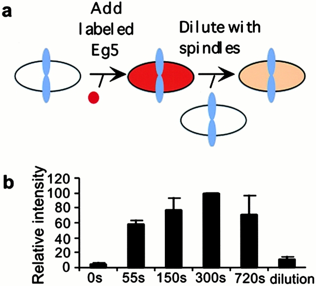
We used fluorescent speckle microscopy to probe the dynamics of the mitotic kinesin Eg5 in Xenopus extract spindles, and compared them to microtubule dynamics. We found significant populations of Eg5 that were static over several seconds while microtubules flux towards spindle poles. Eg5 dynamics are frozen by adenylimidodiphosphate. Bulk turnover experiments showed that Eg5 can exchange between the spindle and the extract with a half life of <55 s. Eg5 distribution in spindles was not perturbed by inhibition of its motor activity with monastrol, but was perturbed by inhibition of dynactin with p50 dynamitin. We interpret these data as revealing the existence of a static spindle matrix that promotes Eg5 targeting to spindles, and transient immobilization of Eg5 within spindles. We discuss alternative interpretations of the Eg5 dynamics we observe, ideas for the biochemical nature of a spindle matrix, and implications for Eg5 function.


Figures

Figure 1.
Figure 1.
Characterization of recombinant Eg5. (a) A chromatograph for the purification by gel filtration of labeled full length Eg5 eluted from nickel charged resin. The absorbance at 280 nm (total protein, blue trace) and 590 nm (Texas red, brown trace) wavelengths of light are shown. Standards with Stokes radii 8.5 and 6.1 nm elute at volumes indicated by arrows, s1 and s2, respectively. (b) Coomassie-stained, labeled full-length Eg5 from the fraction corresponding to the major peak (13.5-nm Stokes radius) by gel filtration chromatography is resolved by SDS-PAGE. Protein from this fraction was directly used in the microscopy experiments. (c) In vitro microtubule gliding velocities of labeled and unlabeled Eg5 in 1 mM ATP. Motility assays were performed as described (Mayer et al., 1999). (d) A spindle prepared for fluorescent speckle microscopy of labeled Eg5 (green). Alexa 488–labeled tubulin and Hoechst were added to the extract to visualize the microtubules (red) and DNA (blue). Fig. 2 c shows the distribution of Eg5 alone in this spindle and Fig. 7 a provides a line scan across the image showing the distribution of Eg5 in the spindle relative to microtubules. Bar, 5 μm.
Figure 7.
Figure 7.
Distribution of Eg5 (green trace) relative to microtubules (red trace) in perturbed and unperturbed spindles. (a) Linescan of fluorescence intensity (arbitrary units) across the control bipolar spindle shown in Fig. 1 d. (b) Linescan across the spindle shown in Fig. 5 showing the distribution of Eg5 in the presence of 100 μM monastrol. (c) Linescan across a bipolar spindle assembled in the presence of p50 dynamitin (Fig. 6).
Figure 2.
Figure 2.
Comparison of tubulin and Eg5 dynamics in spindles by fluorescent speckle microscopy. (a) Fluorescent speckle microscopy of labeled tubulin in spindles assembled in Xenopus egg extracts. The highlighted region across the spindle was used for preparing the kymograph shown in panel b. The angled streaks reveal polewards microtubule movement. (c) Fluorescent speckle microscopy of labeled Eg5 in the spindle. The highlighted region was used to prepare the kymograph shown in panel d. The vertical streaks reveal static Eg5 populations in spindles. (e) The histogram shows the distribution of rates of polewards movement of tubulin and Eg5 speckles. Videos relating to this figure are available at http://www.jcb.org/content/vol154/issue6. Bars, 5 μm.
Figure 3.
Figure 3.
Persistence of Eg5 speckles in spindles is sensitive to a nonhydrolyzable ATP analogue, AMPPNP. (a) The distribution of labeled Eg5 in a bipolar spindle in the presence of 1.5 mM AMPPNP. The highlighted region was used to prepare the kymograph shown in panel b. The vertical streaks correspond to stationary speckles, almost all of which persist for >200 s. Bars, 5 μm.
Figure 4.
Figure 4.
Eg5 exchanges between spindles and the bulk cytoplasm with a half-life < 55 s. (a) A schematic of a pulse-chase experiment to examine the bulk turnover of Eg5 in in vitro spindles. (b) Labeled tubulin was incorporated into spindles assembled in cell-free extract. Texas red–labeled Eg5 was added and after fixed timepoints samples were removed and diluted into fix. The ratio of Eg5 signal in spindles to that of labeled tubulin at each time point after Eg5 addition was determined. Data from three independent experiments is shown. Note saturation labeling by 300 s. Spindle-associated, labeled Eg5 can be diluted away when spindles to which labeled Eg5 has been added to saturation are mixed with 5 vol of unlabeled spindles. The ratio of labeled Eg5 to labeled tubulin, 300 s after mixing spindles with and without labeled Eg5, is shown in the column labeled “dilution”.
Figure 5.
Figure 5.
Inhibition of Eg5 motility does not accumulate the motor at spindle poles. (a) 100 μM monastrol was added to assembled bipolar spindles and a thin (10-μm thick) sample was prepared for microscopy. An overlay showing the distribution of labeled Eg5 (green), tubulin (red), and DNA (blue) in a bipolar spindle 15 min after monastrol addition. (b) Eg5 alone. Linescans comparing the distribution of Eg5 and tubulin in this spindle are shown in Fig. 7 b. Bars, 5 μm.
Figure 6.
Figure 6.
Eg5 distribution in spindles depends on dynactin function. (a) An overlay showing the immunolocalization of endogenous Eg5 (green), tubulin (red), and DNA (blue) in spindles assembled in the presence of p50 dynamitin (0.7 mg/ml), an inhibitor of dynactin. (b) Eg5 alone. (c) An overlay showing the distribution of recombinant labeled Eg5 (green), tubulin, and DNA in spindles assembled in the presence of the dynactin inhibitor. (d) Labeled Eg5 alone. Linescans comparing the distribution of Eg5 and tubulin in spindles assembled in the presence of p50 dynamitin are shown in Fig. 7 c.
Figure 8.
Figure 8.
Two models for the spindle. (a) Eg5 (green). Other motor proteins, including dynein/dynactin (blue), that interact with a static matrix (black) and microtubules (red) that are fluxing polewards (arrows). The nonmicrotubule mechanical ensemble may be an organized structure (black), whose assembly and maintenance can depend on spindle microtubules, or it may be an aspect of spindle cytoarchitecture. (b) A model for the spindle where motors, including Eg5, interact with microtubules alone. These motors crosslink and generate forces against microtubules that flux polewards.

Comment in

  • Searching for a spindle matrix.
    Wells WA. Wells WA. J Cell Biol. 2001 Sep 17;154(6):1102-4. doi: 10.1083/jcb.200108139. J Cell Biol. 2001. PMID: 11564751 Free PMC article.

References

    1. Alexander, S.P., and C.L. Rieder. 1991. Chromosome motion during attachment to the vertebrate spindle: initial saltatory-like behavior of chromosomes and quantitative analysis of force production by nascent kinetochore fibers. J. Cell Biol. 113:805–815. - PMC - PubMed
    1. Blangy, A., H.A. Lane, P. d'Herin, M. Harper, M. Kress, and E.A. Nigg. 1995. Phosphorylation by p34cdc2 regulates spindle association of human Eg5, a kinesin-related motor essential for bipolar spindle formation in vivo. Cell. 83:1159–1169. - PubMed
    1. Blangy, A., L. Arnaud, and E.A. Nigg. 1997. Phosphorylation by p34cdc2 protein kinase regulates binding of the kinesin-related motor HsEg5 to the dynactin subunit p150. J. Biol. Chem. 272:19418–19424. - PubMed
    1. Desai, A., A. Murray, T.J. Mitchison, and C.E. Walczak. 1999. The use of Xenopus egg extracts to study mitotic spindle assembly and function in vitro. Methods Cell Biol. 61:385–412. - PubMed
    1. Dionne, M.A., L. Howard, and D.A. Compton. 1999. NuMA is a component of an insoluble matrix at mitotic spindle poles. Cell Motil. Cytoskeleton. 42:189–203. - PubMed

Publication types

MeSH terms