. 2001 Sep 17;154(6):1197-208.
doi: 10.1083/jcb.200105138.

Vps34p differentially regulates endocytosis from the apical and basolateral domains in polarized hepatic cells

Affiliations

Vps34p differentially regulates endocytosis from the apical and basolateral domains in polarized hepatic cells

P L Tuma et al. J Cell Biol. .

Abstract

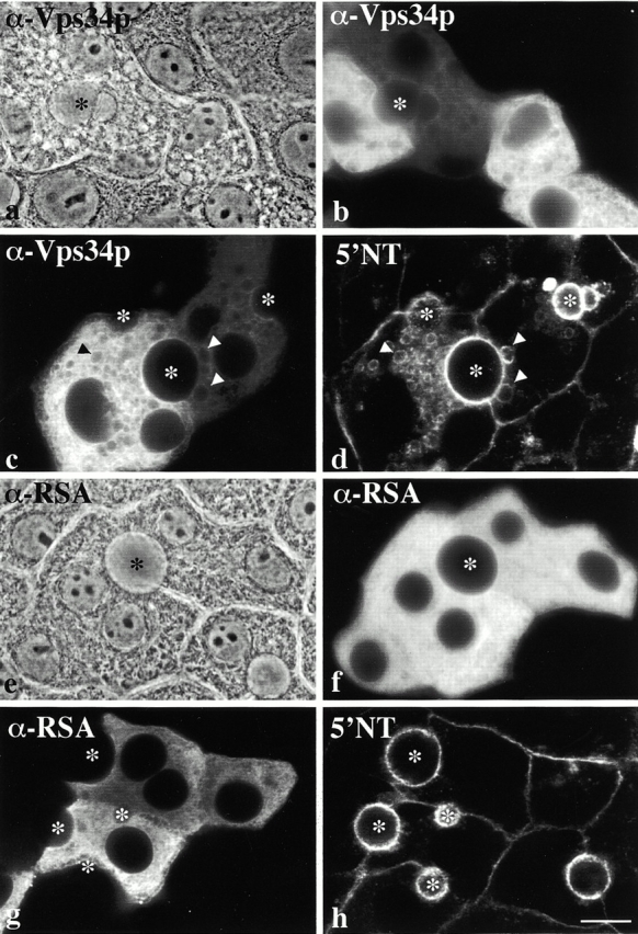
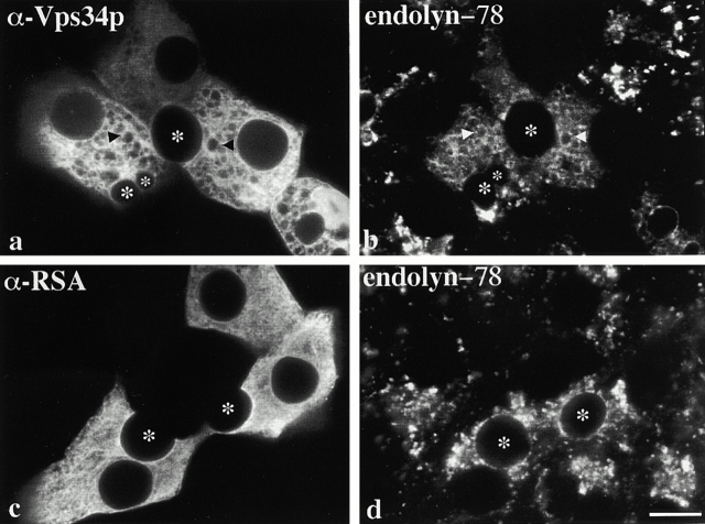
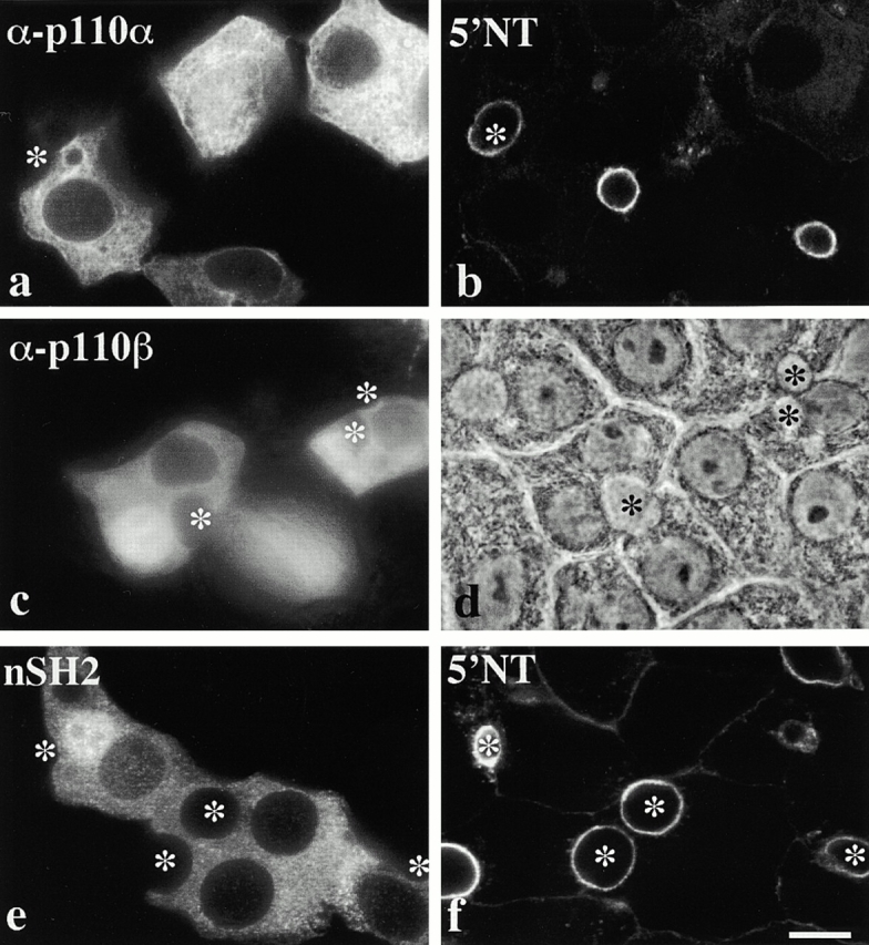
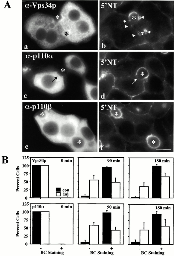
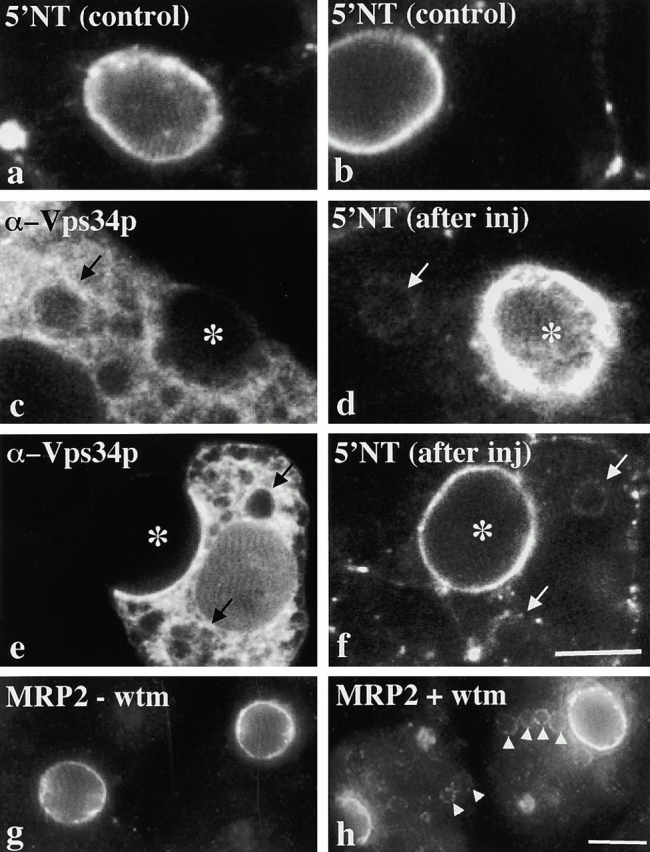
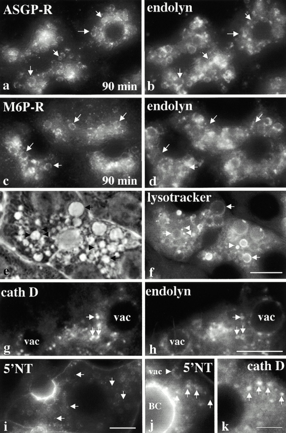
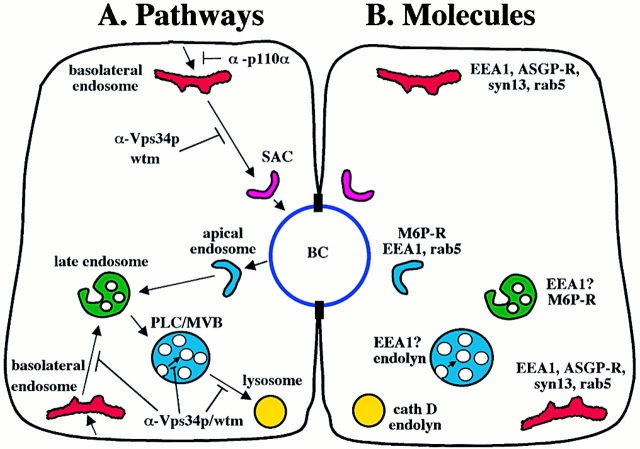
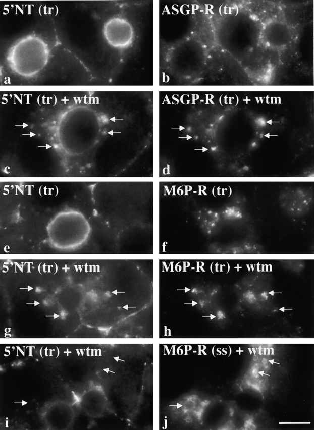
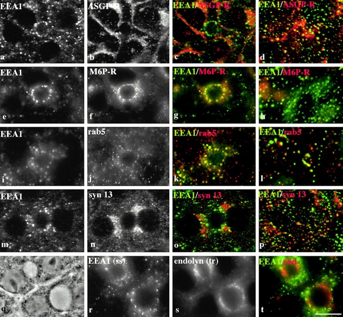
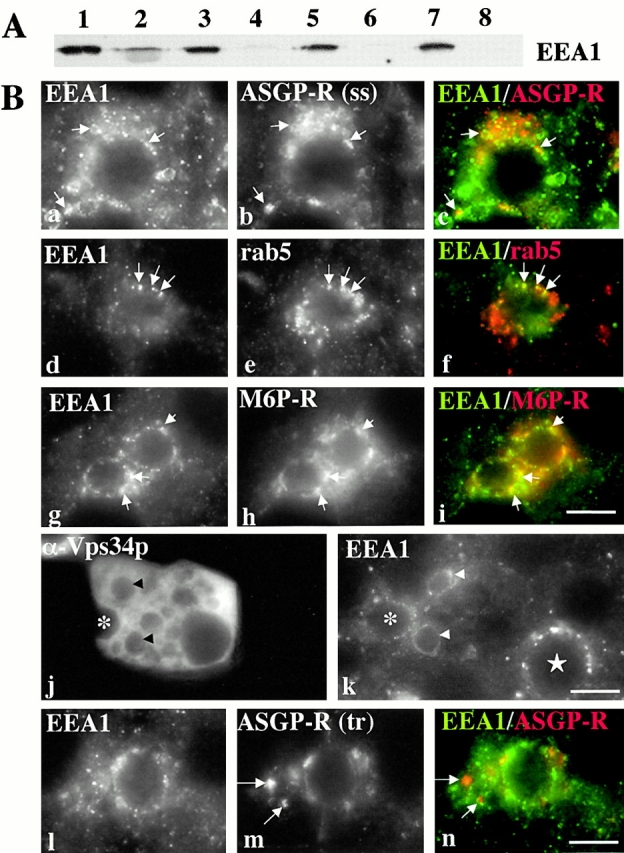
Using a microinjection approach to study apical plasma membrane protein trafficking in hepatic cells, we found that specific inhibition of Vps34p, a class III phosphoinositide 3 (PI-3) kinase, nearly perfectly recapitulated the defects we reported for wortmannin-treated cells (Tuma, P.L., C.M. Finnegan, J.-H Yi, and A.L. Hubbard. 1999. J. Cell Biol. 145:1089-1102). Both wortmannin and injection of inhibitory Vps34p antibodies led to the accumulation of resident apical proteins in enlarged prelysosomes, whereas transcytosing apical proteins and recycling basolateral receptors transiently accumulated in basolateral early endosomes. To understand how the Vps34p catalytic product, PI3P, was differentially regulating endocytosis from the two domains, we examined the PI3P binding protein early endosomal antigen 1 (EEA1). We determined that EEA1 distributed to two biochemically distinct endosomal populations: basolateral early endosomes and subapical endosomes. Both contained rab5, although the latter also contained late endosomal markers but was distinct from the transcytotic intermediate, the subapical compartment. When PI3P was depleted, EEA1 dissociated from basolateral endosomes, whereas it remained on subapical endosomes. From these results, we conclude that PI3P, via EEA1, regulates early steps in endocytosis from the basolateral surface in polarized WIF-B cells. However, PI3P must use different machinery in its regulation of the apical endocytic pathway, since later steps are affected by Vps34p inhibition.


Figures

Figure 1.
Figure 1.
Injection of anti-Vps34p induces formation of vacuoles that contain apical PM proteins. WIF-B cells were injected with anti-Vps34p (a–d) or anti-RSA (e and f). 4 h postinjection, cells were labeled for the injected antibodies (b, c, f, and g) or 5′NT (d and h). Phase micrographs are shown in panels a and e. Arrowheads point to vacuoles containing 5′NT (d and h). Asterisks mark bile canaliculi (BCs) of injected cells. The data are representative of four experiments. Bar, 10 μm.
Figure 2.
Figure 2.
Lysosomal membrane proteins accumulate in anti-Vps34p–induced vacuoles. WIF-B cells were injected with anti-Vps34p (a and b) or anti-RSA (c and d). 4 h postinjection, cells were double-labeled for the injected antibodies (a and c) and endolyn-78 (b and d). Arrowheads point to vacuoles containing endolyn-78. Asterisks mark BCs of injected cells. The data are representative of three experiments. Bar, 10 μm.
Figure 3.
Figure 3.
WIF-B morphology and apical PM protein distributions do not change in cells injected with anti-p110α, anti-p110β, or nSH2 domains. WIF-B cells were injected with anti-p110α (a and b) or anti-p110β (c and d) or nSH2 domains (e and f). 4 h postinjection, cells were labeled for the injected antibodies (a and c), the fusion protein (e), or 5′NT (b and f). The phase micrograph for the anti-p110β–injected cells is shown (d). Asterisks mark BCs of injected cells. The data are representative of at least three experiments. Bar, 10 μm.
Figure 4.
Figure 4.
Anti-Vps34p and anti-p110α impair basolateral to apical transcytosis of apical proteins, but transcytosing molecules do not accumulate in vacuoles. (A) Cells were injected with anti-Vps34p (a and b), -p110α (c and d), or -p110β (e and f) and recovered for 2 h. 5′NT molecules present at the basolateral PM were labeled with antibodies for 15 min at 4°C and the antibody–antigen complexes were chased for 3 h at 37°C. The cells were then fixed and permeabilized and the trafficked molecules visualized with secondary antibodies. Injected antibody staining is shown in a, c, and e while staining of the trafficked 5′NT is shown in b, d, and f. Asterisks indicate the BCs of injected cells. Arrowheads in panel b point to the intracellular accumulations of trafficked 5′NT in anti-Vps34p–injected cells, and the arrows in panels c and d point to unlabeled apical PM in anti-p110α–injected cells. (B) Cells were injected and labeled for 5′NT as described in the legend to A, except the antibody–antigen complexes were chased for 0, 90, or 180 min. For each time point, injected cells were scored for the relative intensity of anti-5′NT immunofluorescence (BC Staining) present at the apical PM. Black bars represent the percentages obtained from control, uninjected neighboring cells (con), while the open bars represent injected cells (inj). Only polarized cells were included in the analysis. Approximately 85–180 cells were examined for each time point. Values are expressed as the mean ± SD. Measurements were performed on at least three experiments. Bar, 10 μm.
Figure 5.
Figure 5.
The apical proteins in vacuoles come from the apical PM. (a–f) 5′NT antibody–antigen complexes were chased to the apical PM for 5 h at 37°C. The cells shown in panels c–f were injected with anti-Vps34p and incubated an additional 4 h. In a and b, 5′NT staining in uninjected cells is shown. Cells double-labeled for the injected antibody and trafficked 5′NT are shown in c and e and d and f, respectively. Arrows are pointing to anti-Vps34p– induced vacuoles that contain apical PM proteins after the additional chase. The data are representative of four experiments. (g and h) WIF-B cells were incubated for 3 h in the absence (g) or presence of 100 nM wortmannin (wtm; h) and stained for MRP2. In untreated cells, MRP2 is detected only at the apical PM (g). Arrowheads point to MRP2-positive vacuoles in treated cells (h). Bars, 10 μm.
Figure 6.
Figure 6.
Wortmannin induces formation of distinct vacuole populations, but does not alter mature lysosome morphology. In panels a–d, cells were treated for 90 min with 100 nM wortmannin. The relative distributions of endolyn-78 to ASGP-R (a and b) or M6P-R (c and d) are shown. Arrows in a and b are pointing to structures that only contain ASGP-R, and in c and d that contain only M6P-R. In e–k, cells were treated for 180 min with 100 nM wortmannin. In e and f, 50 nM lysotracker was added in the final 30 min of wortmannin treatment. Lysotracker staining of acidic compartments in live cells is shown in panel f, and the corresponding phase image in panel e. Enlarged images of wortmannin-treated cells double- labeled for endolyn-78 (h) and cathepsin D (cath D, g) are shown. Arrowheads indicate small puncta containing endolyn at the surface and cathepsin D in the lumen. In panels i–k, 50 μM leupeptin was included hourly in the wortmannin incubations to inhibit acid hydrolase activity. Arrows are pointing to small puncta that contain 5′NT in panel i. Arrows in the enlarged, double-labeled images in j and k are pointing to the small 5′NT-positive puncta that also contain cathepsin D. The data are representative of at least three experiments. Bars, (a–f and i) 10 μm; (g and h) 2 μm; and (j and k) 5 μm.
Figure 10.
Figure 10.
Endocytic and transcytotic pathways in polarized hepatic cells. (A) The intermediates of the transcytotic (top of cell) and endocytic intermediates (bottom of cell) are shown with arrows indicating possible transport routes. (B) The specific markers localized to these intermediates are indicated. Only two intermediates in the basolateral-to-apical transcytotic pathway have been identified: the basolateral early endosome and SAC. We propose that basolateral endocytosis proceeds sequentially through four intermediates: basolateral early endosomes, late endosomes, prelysosomes/MVB, (PLC/MVB) and finally, lysosomes. We further propose that apical endocytosis proceeds through an apical early endosome that is distinct from both the basolateral early endosome and the SAC, and converges with the basolateral endocytic pathway in M6P-R– positive late endosomes. The possible transport blocks imposed by wortmannin, anti-Vps34p, or anti-p110α are indicated in A.
Figure 7.
Figure 7.
Transcytosing apical proteins and recycling receptors internalized from the basolateral PM accumulate in basolateral early endosomes in wortmannin-treated cells. WIF-B cells were treated in the absence (a, b, e, and f) or presence (c, d, and g–j) of 100 nM wortmannin for 2 h. 5′NT and ASGP-R (a–c) or 5′NT and M6P-R (e–h) at the basolateral PM were colabeled for 1 h at 37°C in the continued absence or presence of wortmannin. Arrows are pointing to structures containing both trafficked 5′NT and ASGP-R (c and d) or both 5′NT and M6P-R (g and h). In i and j, 5′NT present at the basolateral PM was continuously labeled for 1 h at 37°C in the presence of wortmannin and its distributions were examined relative to M6P-R at steady state. The data are representative of at least three experiments. Bar, 10 μm.
Figure 8.
Figure 8.
EEA1 is localized to endosomes near the apical PM and the cell periphery that are biochemically distinct. WIF-B cells were double- labeled for EEA1 and ASGP-R (a–d), EEA1 and M6P-R (e–h), EEA1 and rab5 (i–l), or EEA1 and syntaxin13 (syn 13; m–p). In a–c, e–g, i–k, and m–o, the images were focused at the BC, whereas in d, h, i, and p, they were focused at the cell periphery. The merged images from a b, e f, i, j, m, and n are shown in c, g, k, and o, respectively. Only the merged images taken at the cell periphery are shown in d, h, i, and p. In panels q–t, EEA1 steady state distributions (r) are shown relative to endolyn-78 molecules that have been trafficked to the SAC (s). The merged image is shown in panel t. The data are representative of at least three experiments. Bar, 10 μm.
Figure 9.
Figure 9.
Wortmannin and injection of anti-Vps34p alter EEA1 distributions. (A) Cytosolic (lanes 1, 3, 5, and 7) and membrane fractions (lanes 2, 4, 6, and 8) were prepared from WIF-B cells treated for 0 (lanes 1 and 2), 30 (lanes 3 and 4), 60 (lanes 5 and 6), or 120 min (lanes 7 and 8) with 100 nM wortmannin. Equal volumes of the supernatant and pelleted fraction were loaded and immunoblotted with anti-EEA1. (B) WIF-B cells were incubated for 90 or 180 min with 100 nM wortmannin (a–f and g–i, respectively). Cells were fixed, permeabilized, and double-labeled for EEA1 and ASGP-R (a and b), rab5 (d and e), or M6P-R (g and h). Merged images are shown in c, f, and i. Arrowheads point to structures containing both EEA1 and the indicated marker. WIF-B cells were injected with anti-Vps34p, and after 4 h were double-labeled for the injected antibodies (j) and EEA1 (k). Images were focused at slightly different focal planes to better visualize the injected cell in panel j, and EEA1 vacuolar staining in panel k. Arrowheads point to vacuoles containing EEA1. The asterisk marks the BC of the injected cell and the star marks the BC of an uninjected cell. In panels l and m, cells were treated with 100 nM wortmannin for 120 min. ASGP-R at the basolateral surface was continuously labeled with antibodies for an additional hour in the presence of wortmannin. Trafficked ASGP-R distributions (m) were determined relative to EEA1 at steady state (l). The merged image is shown in n. The data are representative of at least three experiments, except in j and k, which are representative of two experiments. Bar, 10 μm.

Similar articles

Cited by

References

    1. Backer, J.M. 2000. Phosphoinositide 3-kinases and the regulation of vesicular trafficking. Mol. Cell. Biol. Res. Commun. 3:193–204. - PubMed
    1. Barr, V.A., and A.L. Hubbard. 1993. Newly synthesized hepatocyte PM proteins are transported in transcytotic vesicles in the bile duct-ligated rat. Gastroenterology. 105:554–571. - PubMed
    1. Barr, V.A., L.J. Scott, and A.L. Hubbard. 1995. Immunoadsorption of hepatic vesicles carrying newly synthesized dipeptidyl peptidase IV and polymeric IgA receptor. J. Biol. Chem. 46:27834–27844. - PubMed
    1. Bomsel, M., K. Prydz, R.G. Parton, J. Gruenberg, and K. Simons. 1989. Endocytosis in filter-grown Madin-Darby Canine Kidney cells. J. Cell Biol. 109:3243–3258. - PMC - PubMed
    1. Brown, W.J., D.B. DeWald, S.D. Emr, H. Plutner, and W.E. Balch. 1995. Role for phosphatidylinositol 3-kinase in the sorting and transport of newly synthesized lysosomal enzymes in mammalian cells. J. Cell Biol. 130:781–796. - PMC - PubMed

Publication types

MeSH terms