. 2001 Oct;21(20):6768-81.
doi: 10.1128/MCB.21.20.6768-6781.2001.

Down-regulation of beta-catenin by activated p53

Affiliations

Down-regulation of beta-catenin by activated p53

E Sadot et al. Mol Cell Biol. 2001 Oct.

Abstract

beta-Catenin is a cytoplasmic protein that participates in the assembly of cell-cell adherens junctions by binding cadherins to the actin cytoskeleton. In addition, it is a key component of the Wnt signaling pathway. Activation of this pathway triggers the accumulation of beta-catenin in the nucleus, where it activates the transcription of target genes. Abnormal accumulation of beta-catenin is characteristic of various types of cancer and is caused by mutations either in the adenomatous polyposis coli protein, which regulates beta-catenin degradation, or in the beta-catenin molecule itself. Aberrant accumulation of beta-catenin in tumors is often associated with mutational inactivation of the p53 tumor suppressor. Here we show that overexpression of wild-type p53, by either transfection or DNA damage, down-regulates beta-catenin in human and mouse cells. This effect was not obtained with transcriptionally inactive p53, including a common tumor-associated p53 mutant. The reduction in beta-catenin level was accompanied by inhibition of its transactivation potential. The inhibitory effect of p53 on beta-catenin is apparently mediated by the ubiquitin-proteasome system and requires an active glycogen synthase kinase 3beta (GSK3beta). Mutations in the N terminus of beta-catenin which compromise its degradation by the proteasomes, overexpression of dominant-negative DeltaF-beta-TrCP, or inhibition of GSKbeta activity all rendered beta-catenin resistant to down-regulation by p53. These findings support the notion that there will be a selective pressure for the loss of wild-type p53 expression in cancers that are driven by excessive accumulation of beta-catenin.


Figures

FIG. 1
FIG. 1
Effect of DOX-induced p53 expression on β-catenin organization and level. MEF cells were plated on coverslips, treated with 5 μg of DOX per ml for 24 h, fixed, and double stained for p53 using an anti-mouse p53 polyclonal antibody (A, C, E, and G) and for β-catenin (β-cat) using a monoclonal anti-β-catenin antibody (B, D, F, and H). Bar, 10 μm.
FIG. 2
FIG. 2
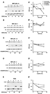
Effect of p53 elevation on β-catenin levels in the Triton X-100-soluble and -insoluble fractions of different cell types. Cells were treated with 5 μg of DOX per ml (A and B) or 5 μg of cisplatin per ml (C and D) for the indicated time periods. Proteins were fractionated into Triton X-100-soluble (lanes s) and -insoluble (lanes i) fractions, resolved by sodium dodecyl sulfate-polyacrylamide gel electrophoresis, and subjected to Western blot analysis using the antibodies indicated in the legend to Fig. 1 and with an antivinculin (vin) antibody. The intensities of the bands from representative gels of MEF p53+/+ (A and C) and MEF p53−/− (B and D) cells were quantified and plotted (A′, C′, B′, and D′, respectively). Extracts from WI38 cells (E and E′) were blotted with the monoclonal anti-β-catenin antibody (β-cat), vinculin (vin), and a mixture of the anti-human p53 antibodies DO1 and 1801 and analyzed as described for MEF cells. a.u., arbitrary units.
FIG. 3
FIG. 3
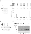
Reciprocal effects of p53 and β-catenin overexpression on the levels of these proteins and the transcriptional activity of β-catenin. (A) p53 or a control empty vector was transfected into 293 cells. After 20 h, cells were fractionated into Triton X-100-soluble (lanes s) and -insoluble (lanes i) fractions and subjected to Western blot analysis. The positions of β-catenin (β-cat), p53, and vinculin (vin) are marked. (B) Transactivation assays with reporter plasmids expressing luciferase under the transcriptional control of different promoters in the presence of p53 and β-catenin. Bars 1 and 2, cyclin G (CycG) promoter; bars 3 and 4, cytomegalovirus promoter; bars 5 to 8, FOPFLASH; bars 9 to 12, TOPFLASH. Cells were collected 20 h after transfection and subjected to luciferase and β-galactosidase assays. The standard error is indicated. (C) Effect of transfected p53 on the levels of cotransfected HA–β-catenin or HA-plakoglobin (PG) blotted with anti-HA antibody. The positions of β-catenin, plakoglobin, and p53 are indicated. (D) H1299 cells (which are p53 deficient) were transfected with increasing amounts of HA–β-catenin (0 to 4 μg) and a constant amount of p53 (100 ng) (lanes 1 to 3) or with a constant amount of HA–β-catenin (4 μg) and increasing amounts of p53 (0 to 600 ng) (lanes 4 to 7). The levels of HA–β-catenin (β-cat) and p53 were determined by Western blot analysis. The endogenous vinculin served as loading control.
FIG. 4
FIG. 4
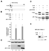
p53 mutants fail to reduce β-catenin (β-cat) levels and transactivation capacity. (A) Schematic representation of the p53Δ13–52 mutant. TAD, transactivation domain. (B) Transactivation in 293 cells, using the TOPFLASH reporter plasmid transfected either alone (bar 1) or with HA–β-catenin (bars 2 to 4) in the presence of wt mouse p53 (bar 3) or the mutant Δ13–52 mouse p53 (bar 4). Cells were harvested 20 h after transfection and subjected to luciferase activity assay. The standard error is indicated. (C) Western blot analysis for β-catenin, p53, and p53Δ13–52 in cell lysates from the experiment in panel B. (D) Human wt p53 or a human p53R175H mutant was cotransfected with HA–β-catenin into 293 cells and subjected to Western blot analysis. (E) p53 can reduce the levels of β-catenin in Mdm2-deficient MEF. p53−/− Mdm2−/− double-mutant MEF were transfected with 5 μg of β-catenin plasmid in the presence or absence of 300 ng of p53 plasmid. The level of the transfected (HA-tagged) β-catenin was determined by Western blot analysis. The positions of β-catenin and p53 are indicated. The asterisks in panels C, D, and E represent a nonspecific band obtained with the anti-HA antibody.
FIG. 5
FIG. 5
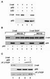
Blocking of GSK3β activity, polyubiquitination, and proteasomal degradation inhibit the effect of p53 on β-catenin (β-cat). (A) wt HA–β-catenin was transfected into 293 cells with p53 (lanes 3 and 4) or without p53 (lanes 1 and 2), and half of the cultures were treated overnight with 30 mM LiCl (lanes 2 and 4) before harvesting of the cells and determination of the levels of HA–β-catenin and p53 by Western blot analysis. (B) wt β-catenin (lanes 1 to 4) or the HA-S33Y β-catenin mutant (lanes 5 to 8) was cotransfected with p53 into 293 cells. After 16 h, MG132 (25 μM) was added to the indicated samples (lanes 3, 4, 7, and 8). The cells were harvested 4 h later and subjected to Western blot analysis. The upper panel shows the levels of β-catenin (wt or S33Y mutant). The lower panel shows the transfected p53. (C) VSV-tagged β-catenin (1 μg) was transfected alone (lane 1) or cotransfected with p53 (1 μg) (lane 2) and increasing concentrations (2 μg [lane 3] and 4 μg [lane 4]) of the dominant-negative HA-tagged ΔF-β-TrCP. The transfected wt β-catenin was detected by anti-VSV antibody, while ΔF-β-TrCP expression was monitored with an anti-HA tag antibody. The asterisk indicates a nonspecific band obtained with the anti-HA antibody.
FIG. 6
FIG. 6
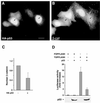
wt p53 down-regulates β-catenin in SW480 CRC cells. (A and B) SW480 cells were transfected with HA-p53 (4 μg) and immunostained for HA (A) and β-catenin (β-cat) (B) 20 h after transfection. The arrows point to the p53-transfected cells. (C) Computerized quantitation of nuclear β-catenin in control, nontransfected cells and in p53-transfected cells. (D) Transactivation in SW480 cells cotransfected with p53 and either TOPFLASH or FOPFLASH reporter plasmid. Cells were harvested 20 h after transfection and subjected to luciferase assay and Western blot analysis for the transfected HA-p53. The position of HA-p53 on the Western blot is indicated. Bar, 10 μm.
FIG. 7
FIG. 7
p53 fails to affect the mutant β-catenin ΔS45 of HCT116 CRC cells. (A to D) HCT116 cells were cultured on coverslips for 24 h in either the presence (A and B) or the absence (C and D) of 5 μg of DOX per ml. After fixation, the cells were double immunostained for p53 using a mixture of DO1 and 1801 antibodies (A and C) and for β-catenin (β-cat) (B and D) using a polyclonal anti-β-catenin antibody. Bar, 10 μm. (E) HCT116 cells were treated with 5 μg of DOX per ml for the indicated times and fractionated into Triton X-100-soluble (lanes s) and -insoluble (lanes i) fractions. The lysates were subjected to Western blot analysis. The positions of β-catenin, p53, and vinculin (vin) (as a control) are indicated. (F) Quantitative analysis of the intensities of the bands shown in panel E. au, arbitrary units.
FIG. 8
FIG. 8
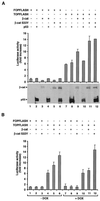
p53 does not inhibit transactivation by the ΔS45 β-catenin of HCT116 cells and of transfected S33Y but inhibits the activity of wt β-catenin in these cells. (A) HCT116 cells were transfected with FOPFLASH (lanes 1 to 5) or TOPFLASH (lanes 7 to 12), together with p53 (lanes 2, 4, 6, 8, 10, and 12), HA–β-catenin (lanes 3, 4, 9, and 10), or p53 and HA-S33Y β-catenin (lanes 5, 6, 11, and 12). Luciferase activity was determined 20 h after transfection, and Western blot analyses of the cell lysates for the expression of β-catenin (β-cat) (wt) and the mutant (S33Y) and for p53 were performed. (B) HCT116 cells were transfected with wt and S33Y β-catenin and either treated with DOX (to elevate p53 levels) as described for Fig. 7 (bars 7 to 12) or left untreated (bars 1 to 6). Transactivation driven by the FOPFLASH and TOPFLASH reporter plasmids was determined. The standard error is shown.

Similar articles

Cited by

References

    1. Abarzua P, LoSardo J E, Gubler M L, Neri A. Microinjection of monoclonal antibody PAb421 into human SW480 colorectal carcinoma cells restores the transcription activation function to mutant p53. Cancer Res. 1995;55:3490–3494. - PubMed
    1. Adams C, Nelson W. Cytomechanics of cadherin-mediated cell-cell adhesion. Curr Opin Cell Biol. 1998;10:572–577. - PubMed
    1. Albrechtsen N, Dornreiter I, Grosse F, Kim E, Wiesmuller L, Deppert W. Maintenance of genomic integrity by p53: complementary roles for activated and non-activated p53. Oncogene. 1999;18:7706–7717. - PubMed
    1. Ashcroft M, Taya Y, Vousden K H. Stress signals utilize multiple pathways to stabilize p53. Mol Cell Biol. 2000;20:3224–3233. - PMC - PubMed
    1. Ashcroft M, Vousden K H. Regulation of p53 stability. Oncogene. 1999;18:7637–7643. - PubMed

Publication types

MeSH terms