. 2001 Oct;21(20):6820-32.
doi: 10.1128/MCB.21.20.6820-6832.2001.

Use of chromatin immunoprecipitation to clone novel E2F target promoters

Affiliations

Use of chromatin immunoprecipitation to clone novel E2F target promoters

A S Weinmann et al. Mol Cell Biol. 2001 Oct.

Abstract

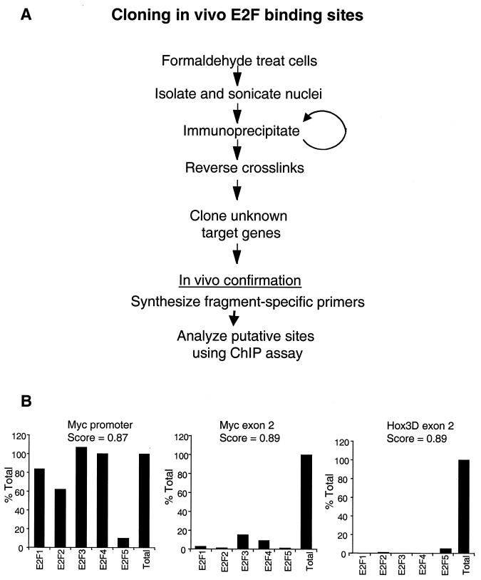
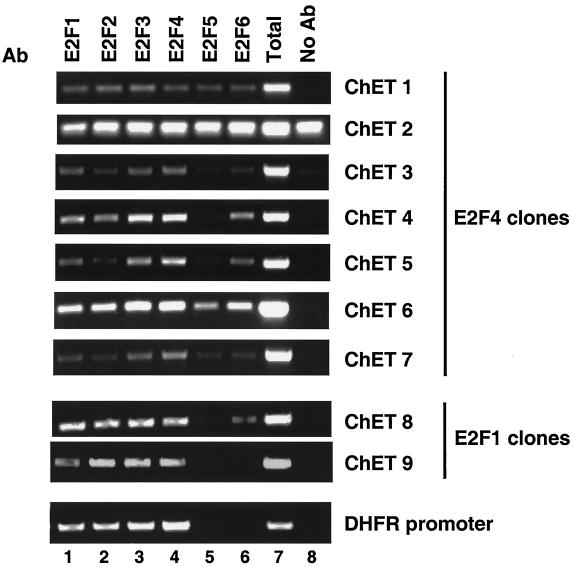
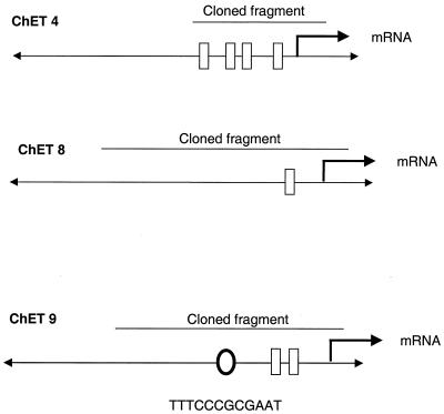
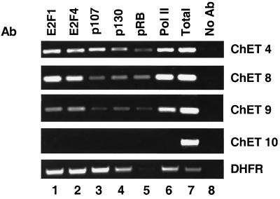
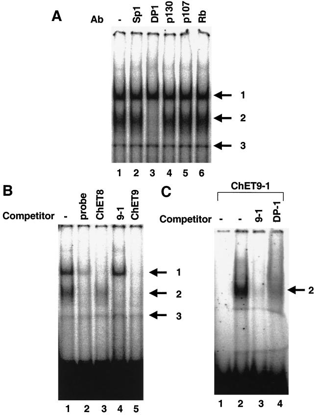
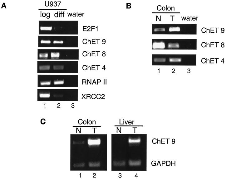
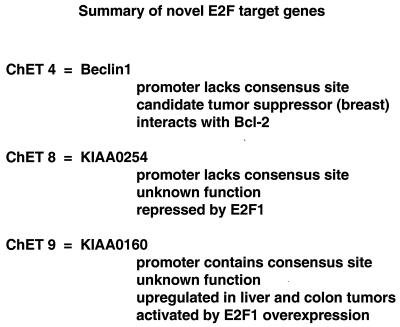
We have taken a new approach to the identification of E2F-regulated promoters. After modification of a chromatin immunoprecipitation assay, we cloned nine chromatin fragments which represent both strong and weak in vivo E2F binding sites. Further characterization of three of the cloned fragments revealed that they are bound in vivo not only by E2Fs but also by members of the retinoblastoma tumor suppressor protein family and by RNA polymerase II, suggesting that these fragments represent promoters regulated by E2F transcription complexes. In fact, database analysis indicates that all three fragments correspond to genomic DNA located just upstream of start sites for previously identified mRNAs. One clone, ChET 4, corresponds to the promoter region for beclin 1, a candidate tumor suppressor protein. We demonstrate that another of the clones, ChET 8, is strongly bound by E2F family members in vivo but does not contain a consensus E2F binding site. However, this fragment functions as a promoter whose activity can be repressed by E2F1. Finally, we demonstrate that the ChET 9 promoter contains a consensus E2F binding site, can be activated by E2F1, and drives expression of an mRNA that is upregulated in colon and liver tumors. Interestingly, the characterized ChET promoters do not display regulation patterns typical of known E2F target genes in a U937 cell differentiation system. In summary, we have provided evidence that chromatin immunoprecipitation can be used to identify E2F-regulated promoters which contain both consensus and nonconsensus binding sites and have shown that not all E2F-regulated promoters show identical expression profiles.


Figures

FIG. 1
FIG. 1
(A) Schematic of the E2F chromatin immunoprecipitation cloning procedure. (B) Graphical representation of the results of a chromatin immunoprecipitation experiment measuring E2F binding at the Myc promoter, Myc exon 2, and Hox3D exon 2. Scores representing homology to the E2F consensus site as determined by computer analysis (21) are shown at the top of each graph. The y axis represents Imagequant quantitation of the amount of specific PCR products expressed as the percentage of antibody binding versus the amount of PCR product obtained using a standarized aliquot of input chromatin. The signal in the no-antibody lane was subtracted from each sample as a nonspecific binding background. The E2F family members used in the immunoprecipitation are shown on the x axis.
FIG. 2
FIG. 2
Confirmation of ChET clones by examining E2F binding in vivo. A representative chromatin immunoprecipitation experiment with HeLa cells is shown. Immunoprecipitation proceeded utilizing antibodies (Ab) against E2F1 (lane 1), E2F2 (lane 2), E2F3 (lane 3), E2F4 (lane 4), E2F5 (lane 5), and E2F6 (lane 6) or no antibody (lane 8). Following DNA purification, samples were subjected to PCR with primers designed for the individual E2F clones or the dhfr promoter as a control (labeled on the right). A portion of the total input was also examined by PCR (lane 7).
FIG. 3
FIG. 3
Genomic organization of the ChET clones. GenBank database searches were performed with the sequences corresponding to ChET 4, ChET 8, and ChET 9. After the locations of the clones were determined, further Blast searches were performed examining the sequences immediately adjacent to the cloned fragments. The locations of adjacent mRNAs are indicated by bent arrows. Positions of consensus Sp1 (rectangles) and E2F (oval) sites are also shown.
FIG. 4
FIG. 4
Characterization of the protein complexes bound in vivo to the ChET clones. A chromatin immunoprecipitation experiment was performed in HeLa cells utilizing antibodies (Ab) to E2F1 (lane 1), E2F4 (lane 2), p107 (lane 3), p130 (lane 4), Rb (lane 5), RNA polymerase II (Pol II; lane 6), or no antibody as a control (lane 8). An aliquot of the total input is also shown (lane 7). Primers to the ChET clones or the DHFR promoter were used in PCRs for analysis.
FIG. 5
FIG. 5
Transient-transfection analysis of ChET promoter-luciferase reporters. (A) A transient-transfection experiment in NIH 3T3 cells was performed with a segment of ChET 8 cloned in either the forward or reverse orientation upstream of luciferase. The y axis of the graph represents the relative luciferase units, with the transfected material shown on the x axis. pGL2 represents the luciferase vector lacking a promoter. (B) A transient-transfection analysis was performed with the ChET 8 promoter-luciferase reporter transfected into NIH 3T3 cells in the presence of 2 μg of an E2F1 expression vector (cytomegalovirus [CMV] E2F1) or the pCDNA3 vector as a control. The cdc2 promoter-luciferase reporter construct was used as a control in both panels A and B. (C) Transient-transfection analysis of the ChET 9-luciferase reporter construct was performed in NIH 3T3 cells. A graphical representation of the results is shown with the dhfr promoter used as a positive control. (D) Overexpression of E2F1 upregulates ChET 9 promoter activity. Cotransfection experiments were performed containing 2 μg of a CMV E2F1 expression construct with the ChET 9-luciferase construct or the dhfr-luciferase reporter vector as a control. Results of the luciferase assay are shown in the graph as indicated for panel A.
FIG. 6
FIG. 6
In vitro EMSA of cloned fragments. In each panel (arrows), complex 3 represents a nonspecific band, complex 2 indicates an E2F-DP complex, and complex 1 indicates a complex containing proteins that have not yet been identified. (A) Supershift EMSA was performed to determine the components of the gel-shifted complexes. Reaction mixtures containing HeLa nuclear extract were incubated with an antibody (Ab) to Sp1 (lane 2), DP-1 (lane 3), p130 (lane 4), p107 (lane 5), or Rb (lane 6) or no antibody (lane 1) followed by incubation with the b-myb E2F site labeled as a probe. (B) EMSA competition experiments using the E2F site from the b-myb promoter as a probe. Oligonucleotides corresponding to the unlabeled probe (lane 2), a fragment spanning the ChET 8 transcription start site (lane 3), an oligonucleotide corresponding to the consensus E2F site from ChET 9 (lane 4), or the ChET 9 fragment (lane 5) were used as competitors. (C) EMSA using the ChET 9 E2F site as a probe. A double-stranded oligonucleotide containing the consensus E2F site within the ChET 9 fragment (the sequence is shown in Fig. 3) was radiolabeled and used for EMSA. The probe was incubated with HeLa nuclear extract and the unlabeled probe (lane 3), a DP-1 antibody (lane 4), or extract alone (lane 2). Lane 1 represents an aliquot of the probe without extract incubation. An arrow to the right of the gel image indicates the specific E2F-DP complex.
FIG. 7
FIG. 7
mRNA expression profiles of the three high-affinity ChET clones. (A) RT-PCR analysis of mRNA expression levels in RNA obtained from either U937 log-phase (lane 1) or differentiated (diff; lane 2) cells. RT-PCR primers complementary to E2F1, ChET 9, ChET 8, ChET 4, RNA polymerase (RNAP) II, or XRCC2 were used as indicated on the right. A water control is shown in lane 3. (B) RT-PCR analysis of RNA from either normal (N) colon (lane 1) or colon tumor (T; lane 2). Primer sets to the specific mRNAs are indicated. (C) RT-PCR analysis of ChET 9 mRNA expression in the RNA obtained from either human normal colon (lane 1), colon tumor (lane 2), normal liver (lane 3), or liver tumor (lane 4). The normal colon and colon tumor samples are the same as those shown in panel B with GAPDH primers added as a loading control.
FIG. 8
FIG. 8
Summary of information obtained relating to the ChET promoter clones.

References

    1. Aita V M, Liang X H, Murty V V, Pincus D L, Yu W, Cayanis E, Kalachikov S, Gilliam T C, Levine B. Cloning and genomic organization of beclin 1, a candidate tumor suppressor gene on chromosome 17q21. Genomics. 1999;59:59–65. - PubMed
    1. Albert T, Wells J, Funk J-O, Pullner A, Raschke E-E, Stelzer G, Meisterernst M, Farnham P J, Eick D. The chromatin structure of the dual c-Myc promoter P1/P2 is regulated by separate elements. J Biol Chem. 2001;276:20482–20490. - PubMed
    1. Boyd K E, Wells J, Gutman J, Bartley S M, Farnham P J. c-Myc target gene specificity is determined by a post-DNA-binding mechanism. Proc Natl Acad Sci USA. 1998;95:13887–13892. - PMC - PubMed
    1. Bussemaker H J, Li H, Siggia E D. Regulatory element detection using correlation with expression. Nat Genet. 2001;27:167–174. - PubMed
    1. Cohen-Kaminsky S, Maouche-Chretien L, Vitelli L, Vinit M-A, Blanchard I, Yamamoto M, Peschle C, Romeo P-H. Chromatin immunoselection defines a TAL-1 target. EMBO J. 1998;17:5151–5160. - PMC - PubMed

Publication types

MeSH terms

LinkOut - more resources