. 2001 Oct;21(20):6841-50.
doi: 10.1128/MCB.21.20.6841-6850.2001.

Growth arrest and DNA damage-inducible protein GADD34 assembles a novel signaling complex containing protein phosphatase 1 and inhibitor 1

Affiliations

Growth arrest and DNA damage-inducible protein GADD34 assembles a novel signaling complex containing protein phosphatase 1 and inhibitor 1

J H Connor et al. Mol Cell Biol. 2001 Oct.

Abstract

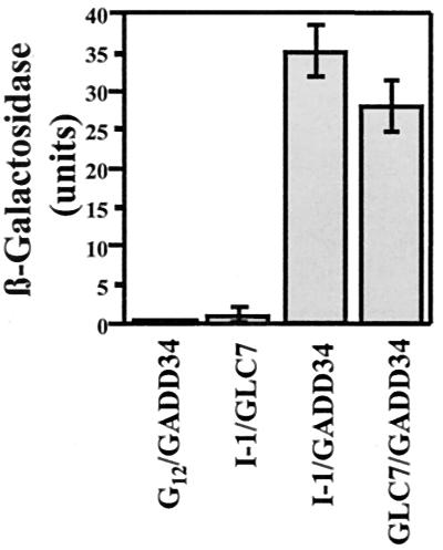
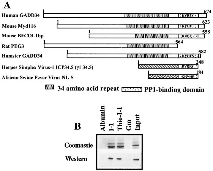
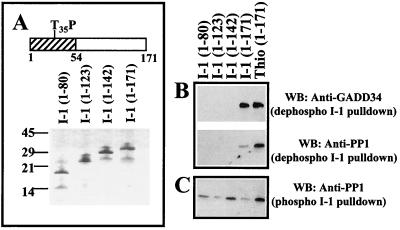
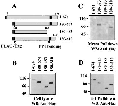
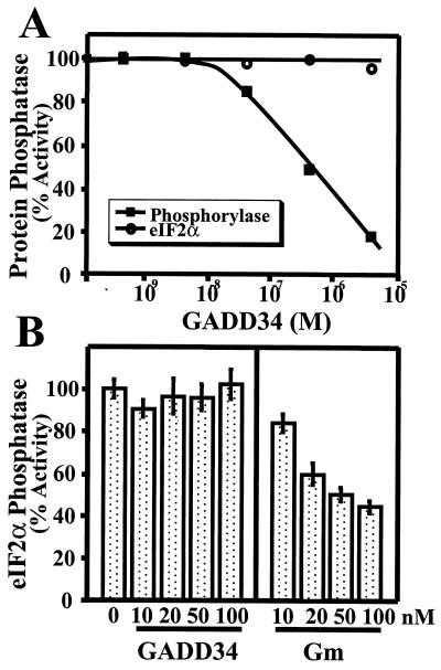
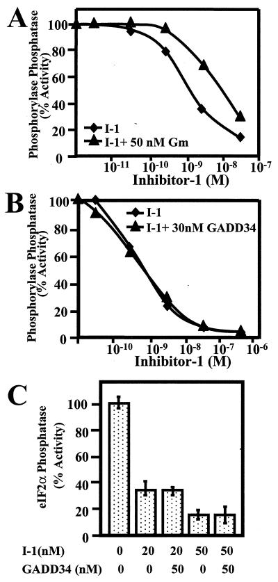
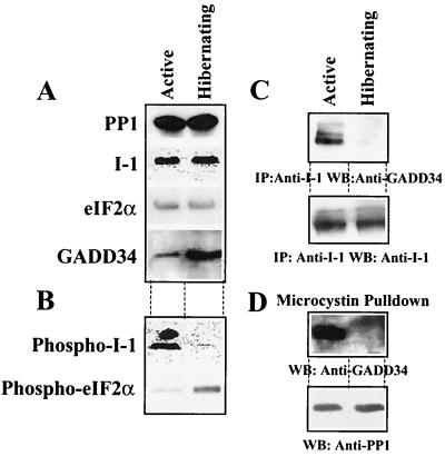
The growth arrest and DNA damage-inducible protein, GADD34, was identified by its interaction with human inhibitor 1 (I-1), a protein kinase A (PKA)-activated inhibitor of type 1 protein serine/threonine phosphatase (PP1), in a yeast two-hybrid screen of a human brain cDNA library. Recombinant GADD34 (amino acids 233 to 674) bound both PKA-phosphorylated and unphosphorylated I-1(1-171). Serial truncations mapped the C terminus of I-1 (amino acids 142 to 171) as essential for GADD34 binding. In contrast, PKA phosphorylation was required for PP1 binding and inhibition by the N-terminal I-1(1-80) fragment. Pulldowns of GADD34 proteins expressed in HEK293T cells showed that I-1 bound the central domain of GADD34 (amino acids 180 to 483). By comparison, affinity isolation of cellular GADD34/PP1 complexes showed that PP1 bound near the C terminus of GADD34 (amino acids 483 to 619), a region that shows sequence homology with the virulence factors ICP34.5 of herpes simplex virus and NL-S of avian sarcoma virus. While GADD34 inhibited PP1-catalyzed dephosphorylation of phosphorylase a, the GADD34-bound PP1 was an active eIF-2alpha phosphatase. In brain extracts from active ground squirrels, GADD34 bound both I-1 and PP1 and eIF-2alpha was largely dephosphorylated. In contrast, the I-1/GADD34 and PP1/GADD34 interactions were disrupted in brain from hibernating animals, in which eIF-2alpha was highly phosphorylated at serine-51 and protein synthesis was inhibited. These studies suggested that modification of the I-1/GADD34/PP1 signaling complex regulates the initiation of protein translation in mammalian tissues.


Figures

FIG. 1
FIG. 1
Identification of GADD34 as a protein interacting with PP1 and I-1. Yeast strains containing pACTII-GADD34 and either pASI-I-1, pAS-GLC7, or control vector pAS-Gi12 were grown on nonselective medium lacking Trp and Leu and selective medium lacking Trp, Leu, Ade, and His as described in the text. The figure shows the strength of the protein-protein interactions as estimated by a liquid β-galactosidase assay, with standard errors.
FIG. 2
FIG. 2
I-1 associates directly with GADD34. (A) GADD34-related proteins, shown schematically with regions of structural and functional homology. These proteins also show structural homology with two viral proteins, HSV-1 ICP34.5 and ASV NL-S, specifically in the C-terminal domains containing putative KIXF PP1-binding motifs. (B) Association of recombinant His-GADD34(230–674) with thiophosphorylated (thio-I-1) and unphosphorylated I-1 covalently linked to Sepharose. Controls included albumin and GST-GM(1–240), the PP1-binding fragment of the skeletal muscle glycogen-targeting subunit, which were also covalently linked to Sepharose. Input shows that almost all GADD34 was bound by immobilized I-1. GADD34 binding was visualized using both Coomassie blue protein stain and Western immunoblotting with anti-GADD34 antibody.
FIG. 3
FIG. 3
Mapping GADD34 binding to I-1. (A) Schematic of I-1 structure, with the N-terminal 54 residues (cross-hatched) previously shown to bind PP1 and the PKA-phosphorylated threonine (T35P) required for PP1 inhibition marked. Polypeptides representing C-terminal truncations of I-1 were expressed in bacteria as hexahistidine-tagged proteins and purified by affinity chromatography on Ni-NTA-agarose. Purity of I-1 proteins is shown by SDS-PAGE on a 10% (wt/vol) polyacrylamide gel stained with Coomassie blue. The I-1 proteins were covalently linked to CNBr-activated Sepharose and used in pulldowns of recombinant His-GADD34(230–674) (B). Pulldowns of PP1 catalytic subunits purified from rabbit skeletal muscle were also undertaken using the immobilized unphosphorylated (B) and phosphorylated I-1 (C). The results are compared with thiophosphorylated I-1 immobilized to Sepharose. I-1-bound proteins were analyzed by SDS-PAGE and Western immunoblotting (WB) with anti-GADD34 and anti-PP1 antibodies.
FIG. 4
FIG. 4
Mapping PP1 and I-1 binding to GADD34. FLAG-tagged GADD34 proteins (schematically shown in panel A with FLAG tag and PP1-binding sites highlighted) were expressed in HEK293T cells. Immunoblotting of total cell extracts with anti-FLAG antibody established relative expression of the GADD34 polypeptides in cells (B). HEK293T cell extracts were incubated with microcystin-LR-Sepharose (labeled Mcyst pulldown) to isolate cellular PP1/GADD34 complexes, and GADD34 was detected by Western immunoblotting (WB) (C). I-1-Sepharose was used to isolate GADD34 from HEK293T cell extracts, and the bound proteins in the “I-1 pulldown” were analyzed by immunoblotting with the anti-HA antibody (D).
FIG. 5
FIG. 5
Modulation of PP1 activity by GADD34. (A) PP1 catalytic subunit isolated from rabbit skeletal muscle was assayed using 32P-labeled phosphorylase a and eIF-2α as substrates in the presence of increasing concentrations of recombinant His-tagged GADD34(230–674). A representative assay from three independent experiments that varied by less than 5% is shown. (B) Effects of increasing concentrations of recombinant human GADD34(230–674) and human GST-GM(1–240) on the in vitro dephosphorylation of eIF-2α by skeletal muscle PP1 catalytic subunit are shown, with standard error bars. The results represent the sum of three independent experiments carried out in duplicate.
FIG. 6
FIG. 6
Effect of GADD34 on PP1 inhibition by I-1. The dose-dependent inhibition of phosphorylase phosphatase activity of the skeletal muscle PP1 catalytic subunit was analyzed in the presence (triangles) and absence (diamonds) of 50 nM recombinant GST-GM(1–240) (A) and 30 nM His-GADD34(230–674) (B). Representative curves from three independent experiments that varied by less than 5% are shown. (C) Inhibition of eIF-2α phosphatase activity of the PP1 catalytic subunit by I-1 in the absence and presence of 50 nM His-GADD34(230–674). Results are the sum of three different experiments carried out in duplicate and are shown with standard error bars.
FIG. 7
FIG. 7
Physiological regulation of PP1/GADD34 and I-1/GADD34 complexes. (A) Comparison of total amounts of PP1 catalytic subunit, I-1, eIF-2α, and GADD34 in extracts from brains of active and hibernating squirrel assessed by Western immunoblotting with the appropriate antibodies. (B) Levels of I-1 phosphorylated on threonine-35 and eIF-2α phosphorylated on serine-51 in brain extracts by immunoblotting with the relevant phosphospecific antibodies. (C) I-1 was immunoprecipitated from squirrel brain extracts using a polyclonal anti-human I-1 antibody. The immunoprecipitates were subjected to SDS-PAGE and blotted with an anti-GADD34 antibody. (D) Microcystin-LR-Sepharose was used to isolate PP1 complexes from squirrel brain extracts. The presence of PP1 and GADD34 in microcystin-LR-bound complexes from brain extracts from active (left lanes) and hibernating (right lanes) ground squirrels was analyzed by immunoblotting with anti-PP1 and anti-GADD34 antibodies, respectively.

Similar articles

Cited by

References

    1. Adler H T, Chinery R, Wu D Y, Kussick S J, Payne J M, Fornace A J, Tkachuk D C. Leukemic HRX fusion proteins inhibit GADD34-induced apoptosis and associate with the GADD34 and hSNF5/INI1 proteins. Mol Cell Biol. 1999;19:7050–7060. - PMC - PubMed
    1. Aggen J B, Nairn A C, Chamberlin R. Regulation of protein phosphatase-1. Chem Biol. 2000;7:13–23. - PubMed
    1. Allen P B, Hvalby O, Jensen V, Errington M L, Ramsay M, Chaudhry F A, Bliss T V, Storm-Mathisen J, Morris R G, Andersen P, Greengard P. Protein phosphatase-1 regulation in the induction of long-term potentiation: heterogeneous molecular mechanisms. J Neurosci. 2000;20:3537–3543. - PMC - PubMed
    1. Bibb J A, Nishi A, O'Callaghan J P, Ule J, Lan M, Snyder G L, Horiuchi A, Saito T, Hisanaga S, Czernik A J, Nairn A C, Greengard P. Phosphorylation of protein phosphatase inhibitor-1 by Cdk5 J. Biol Chem. 2001;276:14490–14497. - PubMed
    1. Blitzer R D, Connor J H, Brown G P, Wong T, Shenolikar S, Iyengar R, Landau E M. Gating of CaMKII by cAMP-regulated protein phosphatase activity during LTP. Science. 1998;280:1940–1942. - PubMed

Publication types

MeSH terms