. 2001 Oct;21(20):6939-50.
doi: 10.1128/MCB.21.20.6939-6950.2001.

Membrane raft-dependent regulation of phospholipase Cgamma-1 activation in T lymphocytes

Affiliations

Membrane raft-dependent regulation of phospholipase Cgamma-1 activation in T lymphocytes

M C Verí et al. Mol Cell Biol. 2001 Oct.

Abstract

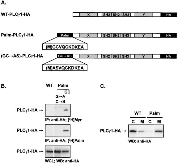
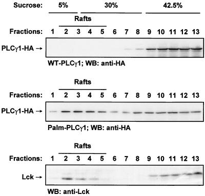
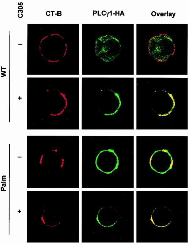
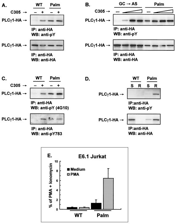
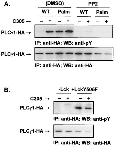
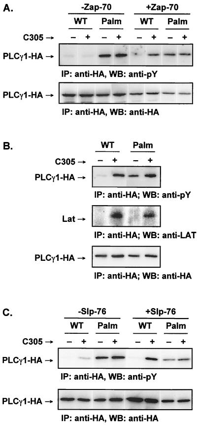
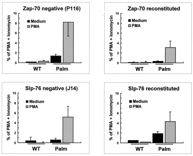
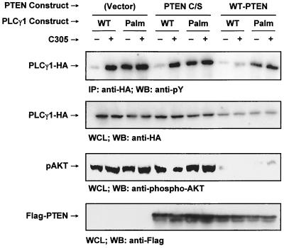
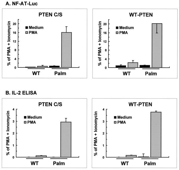
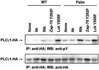
Numerous signaling molecules associate with lipid rafts, either constitutively or after engagement of surface receptors. One such molecule, phospholipase Cgamma-1 (PLCgamma1), translocates from the cytosol to lipid rafts during T-cell receptor (TCR) signaling. To investigate the role played by lipid rafts in the activation of this molecule in T cells, an influenza virus hemagglutinin A (HA)-tagged PLCgamma1 was ectopically expressed in Jurkat T cells and targeted to these microdomains by the addition of a dual-acylation signal. Raft-targeted PLCgamma1 was constitutively tyrosine phosphorylated and induced constitutive NF-AT-dependent transcription and interleukin-2 secretion in Jurkat cells. Tyrosine phosphorylation of raft-targeted PLCgamma1 did not require Zap-70 or the interaction with the adapters Lat and Slp-76, molecules that are necessary for TCR signaling. In contrast, the Src family kinase Lck was required. Coexpression in HEK 293T cells of PLCgamma1-HA with Lck or the Tec family kinase Rlk resulted in preferential phosphorylation of raft-targeted PLCgamma1 over wild-type PLCgamma1. These data show that localization of PLCgamma1 in lipid rafts is sufficient for its activation and demonstrate a role for lipid rafts as microdomains that dynamically segregate and integrate PLCgamma1 with other signaling components.


Figures

FIG. 1
FIG. 1
Characterization and subcellular fractionation of wild-type and acylated PLCγ1 in Jurkat cells. (A) Schematics of the constructs encoding WT-PLCγ1-HA and Palm-PLCγ1-HA. (B) Metabolic labeling of WT-PLCγ1-HA and Palm-PLCγ1-HA. Jurkat TAg cells were transiently transfected with the indicated constructs, labeled with [3H]myristate (top panel) or [3H]palmitate (middle panel), and lysed. Anti-HA precipitates were resolved by SDS-PAGE. A parallel gel loaded with whole-cell lysates (WCL) was immunoblotted (WB) with anti-HA antibody to control for PLCγ1-HA expression (bottom panel). (C) Subcellular fractionation of WT-PLCγ1-HA and Palm-PLCγ1-HA. Jurkat TAg cells were transiently transfected with the indicted constructs and fractionated into cytosol (C) and membrane (M) fractions. A 25-μg amount of protein was resolved by SDS-PAGE and immunoblotted with anti-HA.
FIG. 2
FIG. 2
Lipid raft compartmentalization of wild-type and acylated PLCγ1 in Jurkat cells. Lysates from transiently transfected Jurkat TAg cells were separated on discontinuous sucrose gradients. Fractions were analyzed by SDS-PAGE and immunoblotted (WB) with anti-HA (top and middle panels) or for endogenous Lck (Santa Cruz) (bottom panel).
FIG. 3
FIG. 3
Confocal analysis of the subcellular distribution of wild-type and acylated PLCγ1 in Jurkat cells. Transiently transfected Jurkat TAg cells were activated with an anti-TCR (C305) or left unstimulated as indicated. Cells were stained with CT-B and aggregated with anti-CT-B antibody to visualize lipid rafts (left panels, red). Transfected PLCγ1 was stained with anti-HA (middle panels, green). Areas of colocalization are shown in yellow in overlay images (right panels).
FIG. 4
FIG. 4
Raft-targeted PLCγ1 is constitutively tyrosine phosphorylated and induces NF-AT activation in Jurkat cells. (A) Transfected Jurkat E6.1 cells were activated with an anti-TCR antibody (C305) as indicated. Lysates were immunoprecipitated (IP) with anti-HA, resolved by SDS-PAGE, immunoblotted (WB) with an antiphosphotyrosine 4G10 (anti-pY, upper panel), stripped, and reprobed with anti-HA (lower panel). (B) Jurkat E6.1 cells were transiently transfected with the control GC→AS PLCγ1-HA or Palm-PLCγ1-HA and stimulated with increasing amounts of anti-TCR antibody (C305). Lysates were immunoprecipitated with anti-HA, resolved by SDS-PAGE, immunoblotted with 4G10 (anti-pY, upper panel), stripped, and reprobed with anti-HA (lower panel). (C) Lysates from transfected E6.1 Jurkat cells were immunoprecipitated with anti-HA, resolved by SDS-PAGE, and immunoblotted with 4G10 (anti-pY, upper panel) or a phospho-specific anti-PLCγ1[pY783] (lower panel). (D) The detergent-soluble (S) and raft (R) fractions were separated by discontinuous sucrose gradient, pooled, immunoprecipitated with anti-HA, resolved by SDS-PAGE, immunoblotted with 4G10 (anti-pY, upper panel), stripped, and reprobed with anti-HA (lower panel). (E) Jurkat cells were transiently transfected with the indicated constructs along with an NF-AT luciferase reporter and an adenovirus major late promoter β-galactosidase-encoding vector to control for transfection efficiency. Transfected cells were treated with medium alone, a phorbol ester (PMA), or PMA plus ionomycin. Data were normalized for β-galactosidase activity and expressed as the percentage of maximum activation with PMA plus ionomycin. The data shown are the means and standard errors of the mean of four separate experiments.
FIG. 5
FIG. 5
Tyrosine phosphorylation of raft-targeted PLCγ1 in Jurkat cells requires Lck. (A) Transiently transfected Jurkat E6.1 cells were cultured in medium containing vehicle alone (0.2% dimethyl sulfoxide [DMSO]) or 20 μM PP2. The cells were activated with anti-TCR antibody (C305) as indicated. Lysates were immunoprecipitated (IP) with anti-HA, resolved by SDS-PAGE, immunoblotted (WB) with 4G10 (anti-pY, upper panel), stripped and reprobed with anti-HA (lower panel). (B) The Lck-defective Jurkat cell line JCam1.6 was transfected with Palm-PLCγ1-HA and cotransfected with an empty vector or pSX-LckY505F. Lysates were immunoprecipitated with anti-HA, resolved by SDS-PAGE, immunoblotted with 4G10 (anti-pY, upper panel), stripped, and reprobed with anti-HA (lower panel).
FIG. 6
FIG. 6
Tyrosine phosphorylation of raft-targeted PLCγ1 in Jurkat cells is independent of Zap-70, Lat, and Slp-76. (A) The Zap-70-negative Jurkat line P116 and a stable Zap-70-reconstituted line were transiently transfected and activated as indicated. Lysates were immunoprecipitated (IP) with anti-HA, resolved by SDS-PAGE, immunoblotted (WB) with 4G10 (anti-pY, upper panel), stripped, and reprobed with anti-HA (lower panel). (B) Jurkat E6.1 cells were transfected and activated as indicated. Lysates were immunoprecipitated with anti-HA, resolved by SDS-PAGE, and separately immunoblotted with 4G10 (anti-pY) (top panel), with anti-Lat (middle panel), or with anti-HA (bottom panel). Calf intestinal phosphatase was used to remove phosphate groups from the anti-HA immunoprecipitates probed with anti-Lat to enhance blotting. (C) The Slp-76-deficient Jurkat clone J14 and a stable Slp-76-reconstituted clone (J14-76-11) were transiently transfected and activated as indicated. Lysates were immunoprecipitated with anti-HA, resolved by SDS-PAGE, immunoblotted with 4G10 (anti-pY) (upper panel), stripped, and reprobed with anti-HA (lower panel).
FIG. 7
FIG. 7
NF-AT activation by raft-targeted PLCγ1 does not require Zap-70 or Slp-76. The Zap-70-negative Jurkat line P116, a Zap-70-reconstituted P116 cell-derived line, the Slp-76-deficient Jurkat clone J14, and a Slp-76-reconstituted J14 cell clone (J14-76-11) were transiently transfected with the indicated PLCγ1 constructs along with a NF-AT luciferase reporter and an adenovirus major late promoter β-galactosidase-encoding vector to control for transfection efficiency. Transfected cells were treated with medium alone, PMA, or PMA plus ionomycin. Data were normalized for β-galactosidase activity and expressed as percentage of maximum activation with PMA plus ionomycin. The data shown are the means and standard errors of the mean of three separate experiments.
FIG. 8
FIG. 8
PTEN expression does not affect tyrosine phosphorylation of raft-targeted PLCγ1 in Jurkat T cells. Jurkat TAg cells were transiently transfected with 20 μg of empty vector, Flag-PTEN C/S, or Flag-WT-PTEN along with 20 μg of WT-PLCγ1-HA or Palm-PLCγ1-HA. The cells were treated as indicated. Cell lysates were immunoprecipitated (IP) with anti-HA, resolved by SDS-PAGE, and immunoblotted (WB) with 4G10 (anti-pY) (top panel). Whole-cell lysates (WCL) were immunoblotted with anti-HA (second panel from the top), an anti-phospho-Akt (pAKT, Ser473) (second panel from the bottom), or anti-Flag (bottom panel).
FIG. 9
FIG. 9
PTEN reconstitution does not affect NF-AT activation or IL-2 secretion in Jurkat T cells expressing raft-targeted PLCγ1. (A). Jurkat TAg cells were transiently transfected with 20 μg of Flag-PTEN C/S or Flag-WT-PTEN along with 5 μg of WT-PLCγ1-HA or Palm-PLCγ1-HA, a NF-AT luciferase reporter, and an adenovirus major late promoter β-galactosidase-encoding vector as described in Materials and Methods. Transfected cells were treated with medium alone, PMA, or PMA plus ionomycin. Parallel samples were processed for PLCγ1-HA expression by immunoblot analysis. Data were normalized for PLCγ1-HA expression levels and presented as percentage of maximum activation with PMA plus ionomycin. The data shown are the means and standard error of the mean of four separate experiments. (B) Jurkat TAg cells were transiently transfected with Flag-PTEN C/S or Flag-WT-PTEN along with WT-PLCγ1-HA or Palm-PLCγ1-HA. Transfected cells were treated with medium alone, PMA, or PMA plus ionomycin. Parallel samples were processed for PLCγ1-HA expression by immunoblot analysis. Data were normalized for PLCγ1-HA expression and are presented as a percentage of maximum activation with PMA plus ionomycin. IL-2 secretion by cells treated with PMA plus ionomycin ranged from 2,400 to 3,900 U per 5 × 10⁴ cells. ELISA, enzyme-linked immunosorbent assay.
FIG. 10
FIG. 10
Preferential phosphorylation of raft-targeted PLCγ1 by Rlk and Lck in HEK 293T cells. HEK 293T cells were transiently transfected with 1 μg of PLCγ1-HA and 0.3 μg of the indicated kinase plasmids. Lysates were immunoprecipitated (IP) with anti-HA, resolved by SDS-PAGE, immunoblotted (WB) with 4G10 (anti-pY) (upper panel), stripped, and reprobed with anti-HA (lower panel).

References

    1. Boerth N J, Sadler J J, Bauer D E, Clements J L, Gheith S M, Koretzky G A. Recruitment of SLP-76 to the membrane and glycolipid-enriched membrane microdomains replaces the requirement for linker for activation of T cells in T cell receptor signaling. J Exp Med. 2000;192:1047–1058. - PMC - PubMed
    1. Brown D A, London E. Structure and function of sphingolipid- and cholesterol-rich membrane rafts. J Biol Chem. 2000;275:17221–17224. - PubMed
    1. Chan A C, Dalton M, Johnson R, Kong G H, Wang T, Thoma R, Kurosaki T. Activation of ZAP-70 kinase activity by phosphorylation of tyrosine 493 is required for lymphocyte antigen receptor function. EMBO J. 1995;14:2499–2508. - PMC - PubMed
    1. Crabtree G R, Clipstone N A. Signal transmission between the plasma membrane and nucleus of T lymphocytes. Annu Rev Biochem. 1994;63:1045–1083. - PubMed
    1. DeBell K E, Stoica B A, Veri M C, Di Baldassarre A, Miscia S, Graham L J, Rellahan B L, Ishiai M, Kurosaki T, Bonvini E. Functional independence and interdependence of the src homology domains of phospholipase C-gamma1 in B-cell receptor signal transduction. Mol Cell Biol. 1999;19:7388–7398. - PMC - PubMed

Publication types

MeSH terms

Substances