. 2001 Oct;21(20):7065-77.
doi: 10.1128/MCB.21.20.7065-7077.2001.

The p65 (RelA) subunit of NF-kappaB interacts with the histone deacetylase (HDAC) corepressors HDAC1 and HDAC2 to negatively regulate gene expression

Affiliations

The p65 (RelA) subunit of NF-kappaB interacts with the histone deacetylase (HDAC) corepressors HDAC1 and HDAC2 to negatively regulate gene expression

B P Ashburner et al. Mol Cell Biol. 2001 Oct.

Abstract

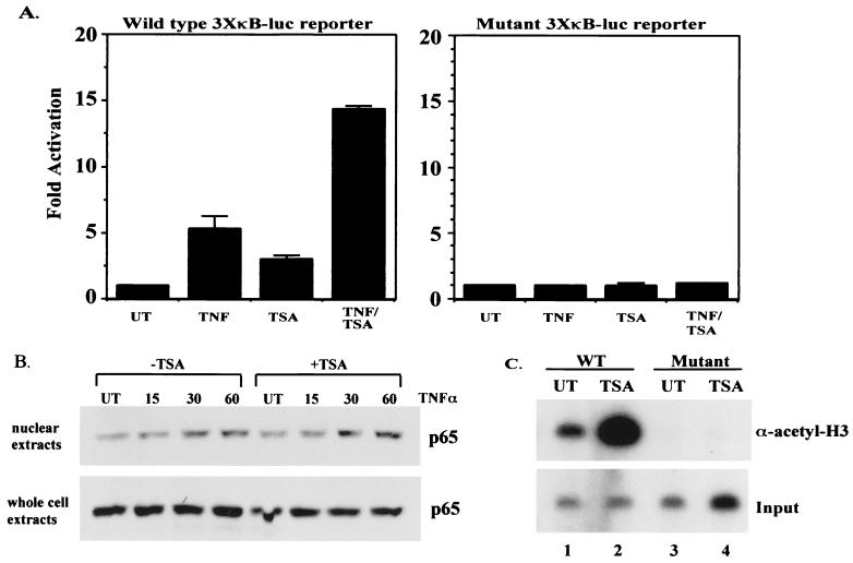
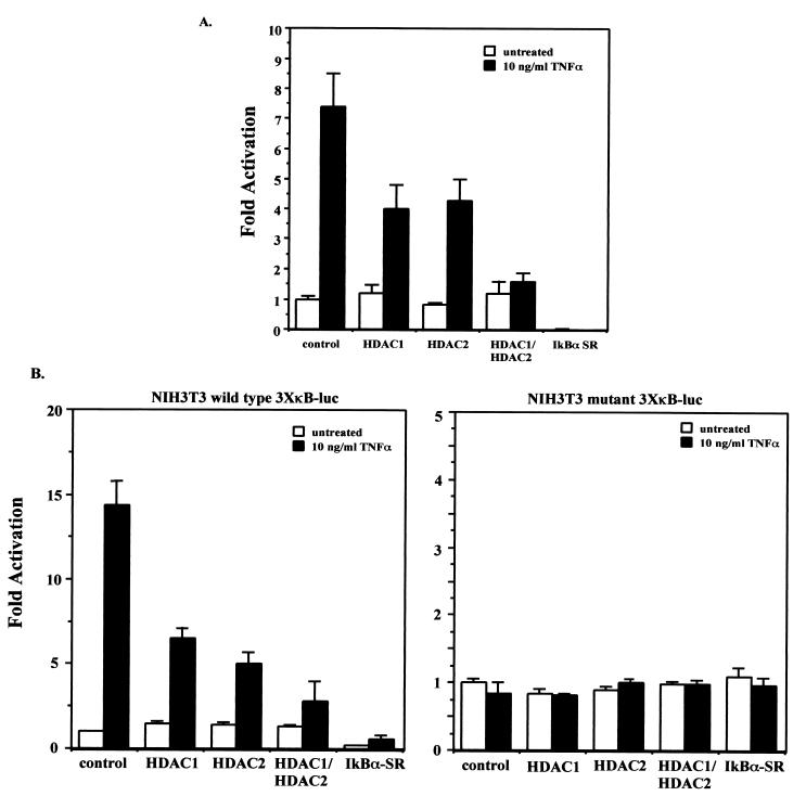
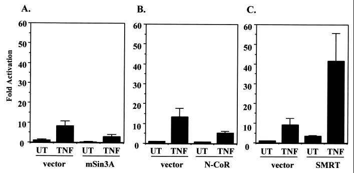
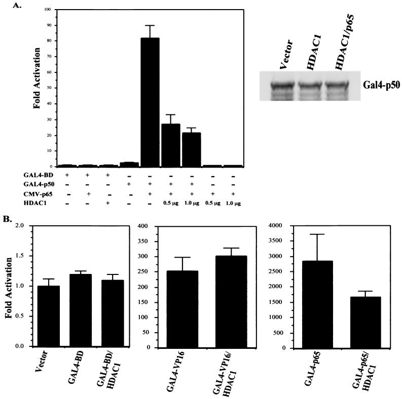
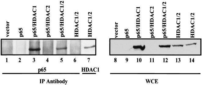
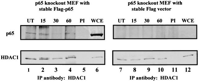
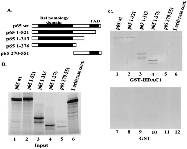
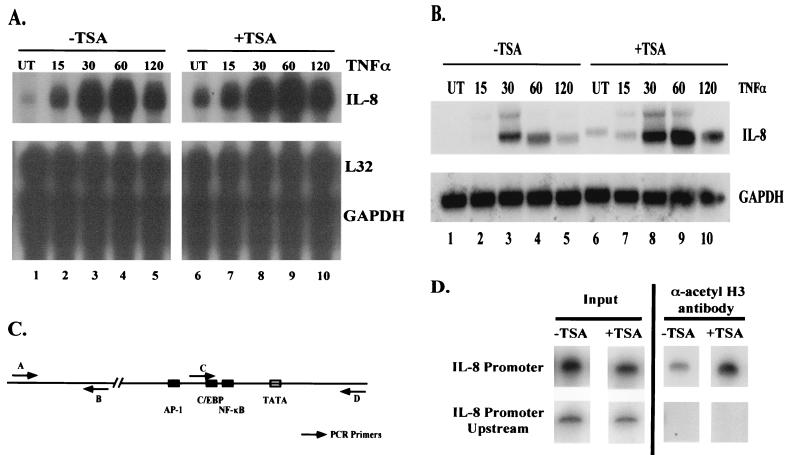
Regulation of NF-kappaB transactivation function is controlled at several levels, including interactions with coactivator proteins. Here we show that the transactivation function of NF-kappaB is also regulated through interaction of the p65 (RelA) subunit with histone deacetylase (HDAC) corepressor proteins. Our results show that inhibition of HDAC activity with trichostatin A (TSA) results in an increase in both basal and induced expression of an integrated NF-kappaB-dependent reporter gene. Chromatin immunoprecipitation (ChIP) assays show that TSA treatment causes hyperacetylation of the wild-type integrated NF-kappaB-dependent reporter but not of a mutant version in which the NF-kappaB binding sites were mutated. Expression of HDAC1 and HDAC2 repressed tumor necrosis factor (TNF)-induced NF-kappaB-dependent gene expression. Consistent with this, we show that HDAC1 and HDAC2 target NF-kappaB through a direct association of HDAC1 with the Rel homology domain of p65. HDAC2 does not interact with NF-kappaB directly but can regulate NF-kappaB activity through its association with HDAC1. Finally, we show that inhibition of HDAC activity with TSA causes an increase in both basal and TNF-induced expression of the NF-kappaB-regulated interleukin-8 (IL-8) gene. Similar to the wild-type integrated NF-kappaB-dependent reporter, ChIP assays showed that TSA treatment resulted in hyperacetylation of the IL-8 promoter. These data indicate that the transactivation function of NF-kappaB is regulated in part through its association with HDAC corepressor proteins. Moreover, it suggests that the association of NF-kappaB with the HDAC1 and HDAC2 corepressor proteins functions to repress expression of NF-kappaB-regulated genes as well as to control the induced level of expression of these genes.


Figures

FIG. 1
FIG. 1
Inhibition of HDAC activity causes an increase in basal and induced expression of an integrated NF-κB-dependent reporter gene (3XκB-Luc). (A) NIH 3T3 cells harboring either integrated wild-type 3XκB-Luc (left) or mutant 3XκB-Luc (right) were treated in triplicate with TNF (10 ng/ml) for 8 h, TSA (100 nM) for 18 h, or with TSA for 18 h and TNF for the final 8 h. Extracts were prepared and relative luciferase activities were determined by normalizing to total protein. Activity is expressed as fold activation relative to the untreated control and is the average ± standard deviation (SD) from a representative experiment. Experiments were performed a minimum of three times, with similar results. UT, untreated cells. (B) Western blot analysis on nuclear and whole-cell extracts probing for p65 levels. (C) ChIP assay. ChIP assays were performed on NIH 3T3 3XκB-Luc wild-type (lanes 1 and 2) and mutant (lanes 3 and 4) cells. Cells were treated with either vehicle (dimethyl sulfoxide) or TSA for 18 h. DNA and protein were cross-linked with formaldehyde, and DNA was sheared and immunoprecipitated with anti-acetyl histone H3 antibody. After reversing cross-links, the DNA was amplified using end-labeled primers specific for the promoter region of the integrated 3XκB-Luc reporter gene. PCR products were analyzed by polyacrylamide gel electrophoresis and bands were visualized by autoradiography. Input, DNA prior to immunoprecipitation with the anti-acetyl histone H3 antibody.
FIG. 2
FIG. 2
(A) HDAC1 and HDAC2 repress TNF-induced expression of a transiently transfected 3XκB-Luc reporter gene. Cos-7 cells were transfected in triplicate with the wild-type 3XκB-Luc reporter and a control vector (pcDNA3.1), pcDNA-HDAC1, pcDNA-HDAC2 alone, or HDAC1 and -2 together. Twenty-four hours after transfection the cells were either untreated or treated with 10 ng of TNF-α/ml for 8 h. Extracts were prepared and relative luciferase activities were determined by normalizing to total protein. Activity is expressed as fold activation relative to the untreated control and is the average ± SD from a representative experiment. Experiments were performed a minimum of three times, with similar results. (B) NIH 3T3 cells harboring either integrated wild-type 3XκB-Luc (left) or mutant 3XκB-Luc (right) were transfected in triplicate as described for panel A. Twenty-four hours after transfection the cells were either untreated or treated with 10 ng of TNF-α/ml for 8 h. Extracts were prepared and relative luciferase activities were determined by normalizing to total protein. Activity is expressed as fold activation relative to the untreated control and is the average ± SD from a representative experiment. Experiments were performed a minimum of three times, with similar results.
FIG. 3
FIG. 3
mSin3a and N-CoR, but not SMRT, can repress TNF-induced expression of 3XκB-Luc. Cos-7 cells were transfected with wild-type 3XκB-Luc and either an appropriate control vector or pIRESHis-mSin3a (A), pCEP4–N-CoR (B), or pCMX-SMRT (C). Twenty-four hours after transfection the cells were either untreated (UT) or treated with 10 ng of TNF-α/ml for 8 h. Extracts were prepared and relative luciferase activities were determined by normalizing to total protein. Activity is expressed as fold activation relative to the untreated control and is the average ± SD from a representative experiment. Experiments were performed a minimum of three times, with similar results.
FIG. 4
FIG. 4
(A) HDAC1 targets the p65 subunit of NF-κB. HeLa cells were transfected with a 5XGAL4-Luc reporter plasmid along with either the GAL4 DNA-binding domain (GAL4-BD) as a control or a full-length fusion of p50 to the GAL4 DNA-binding domain (GAL4-p50). Where indicated, cells were also transfected with pCMV-p65 and/or pcDNA-HDAC1 (either 0.5 or 1.0 μg). Forty-eight hours after transfection extracts were prepared and relative luciferase activities were determined by normalizing to total protein. Activity is expressed as fold activation relative to the untreated control and is the average ± SD from a representative experiment. Experiments were performed a minimum of three times, with similar results. Right panel, Western blot probing for GAL4-p50 to show that its expression was not affected by expression of HDAC1. (B) HeLa cells were transfected with the GAL4 DNA-binding domain, GAL4-VP16, or GAL4-p65 and either control vector or pcDNA-HDAC1 along with 5XGAL4-Luc reporter plasmid. Forty-eight hours after transfection extracts were prepared and relative luciferase activities were determined by normalizing to total protein. Activity is expressed as fold activation relative to the untreated control and is the average ± SD from a representative experiment. Experiments were performed a minimum of three times, with similar results.
FIG. 5
FIG. 5
HDAC1 interacts with p65 in transient transfections. 293 cells were transfected with the indicated plasmids and extracts were made and used for immunoprecipitations (IP) with either a p65-specific antibody (lanes 1 to 6) or an HDAC1-specific antibody (lane 7). Immunoprecipitates were then used in Western blot analysis to probe for the presence of HDAC1. Lanes 8 to 14, Western blot probed for HDAC1 on the whole-cell extracts (WCE) used for the immunoprecipitation reactions.
FIG. 6
FIG. 6
p65 can be coimmunoprecipitated with endogenous HDAC1. Immunoprecipitations (IP) were performed with an antibody specific for HDAC1 on whole-cell extracts (WCE) from p65 null MEFs that had been stably reconstituted with Flag-tagged p65 (left) or the Flag vector (right). Lower panels, Western blot probing the immunoprecipitations for HDAC1 from the Flag-p65-reconstituted cells (lanes 1 to 5) or from the Flag vector-reconstituted cells (lanes 7 to 11); upper panels, Western blot probed for p65 from the HDAC1 immunoprecipitations. Lanes 6 and 12, whole-cell extracts to show the presence of HDAC1 and p65 in the extracts. Prior to harvesting extracts, the cells were either untreated (UT) or treated with 10 ng of TNF-α/ml for the indicated times. PI, preimmune serum.
FIG. 7
FIG. 7
HDAC1 interacts directly with p65 in in vitro binding assays. (A) Diagram of p65 full-length and deletion mutants used for in vitro interaction. (B) Lanes 1 to 6, 1/10 of the indicated input in vitro transcription and translation products used for in vitro binding assays. The products of the in vitro transcription and translation reactions were fractionated by SDS-polyacrylamide gel electrophoresis and the bands were visualized by autoradiography. (C) After the in vitro transcription and translation reactions were performed, 10 μl (of a 50-μl reaction) of the indicated products was mixed with GST-HDAC1 (upper panel) or GST (lower panel) bound to glutathione-agarose beads. After incubation for 1 h at 4°C, the beads were washed extensively and interacting proteins were visualized as for panel A.
FIG. 8
FIG. 8
Inhibition of HDAC activity causes an increase in IL-8 expression. (A) Ribonuclease protection assay showing effect of TSA treatment on IL-8 expression. Total cellular RNA was harvested from HeLa cells after the indicated treatments and used in a ribonuclease protection assay. TNF-α was used at a final concentration of 10 ng/ml for the indicated times. TSA treatment was for 18 h at a final concentration of 100 nM. L32 and GAPDH are shown as loading controls. Other genes used in the assay are not shown. (B) Same as panel A, but RNA was used in Northern blot analysis to analyze the IL-8 expression pattern. (C) Diagram of IL-8 promoter and IL-8 upstream region. Arrows indicate primer pairs used in PCR for ChIP assays. (D) ChIP assay on IL-8 promoter and IL-8 upstream region. Cells were treated with either vehicle (dimethyl sulfoxide) or TSA for 18 h. DNA and protein were cross-linked with formaldehyde, and DNA was sheared and immunoprecipitated with anti-acetyl histone H3 antibody. After reversing cross-links, the DNA was amplified using end-labeled primers specific for the promoter region of the IL-8 gene or for a region upstream of the IL-8 promoter devoid of any known promoter elements. PCR products were analyzed by polyacrylamide gel electrophoresis and bands were visualized by autoradiography. Input, DNA prior to immunoprecipitation with the anti-acetyl histone H3 antibody. UT, untreated cells.

References

    1. Ashburner B P, Shackelford R E, Baldwin A S, Jr, Paules R S. Lack of involvement of ataxia telangiectasia mutated (ATM) in regulation of nuclear factor-kappaB (NF-kappaB) in human diploid fibroblasts. Cancer Res. 1999;59:5456–5460. - PubMed
    1. Ayer D E, Lawrence Q A, Eisenman R N. Mad-Max transcriptional repression is mediated by ternary complex formation with mammalian homologs of yeast repressor Sin3. Cell. 1995;80:767–776. - PubMed
    1. Baldwin A S., Jr The NF-kappa B and I kappa B proteins: new discoveries and insights. Annu Rev Immunol. 1996;14:649–683. - PubMed
    1. Bannister A J, Kouzarides T. The CBP co-activator is a histone acetyltransferase. Nature. 1996;384:641–643. - PubMed
    1. Carlotti F, Dower S K, Qwarnstrom E E. Dynamic shuttling of nuclear factor kappa B between the nucleus and cytoplasm as a consequence of inhibitor dissociation. J Biol Chem. 2000;275:41028–41034. - PubMed

Publication types

MeSH terms