. 2001 Oct;21(20):7097-104.
doi: 10.1128/MCB.21.20.7097-7104.2001.

Mitochondrial import driving forces: enhanced trapping by matrix Hsp70 stimulates translocation and reduces the membrane potential dependence of loosely folded preproteins

Affiliations

Mitochondrial import driving forces: enhanced trapping by matrix Hsp70 stimulates translocation and reduces the membrane potential dependence of loosely folded preproteins

A Geissler et al. Mol Cell Biol. 2001 Oct.

Abstract

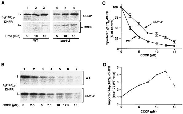
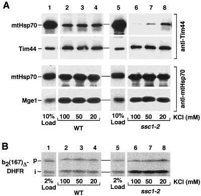
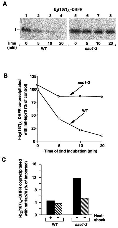
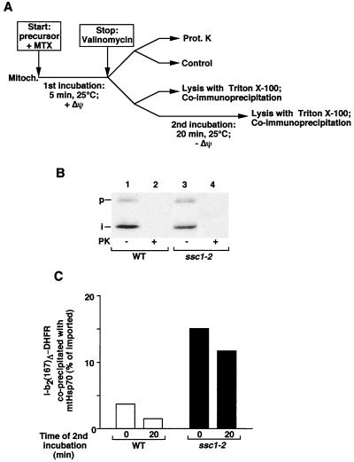
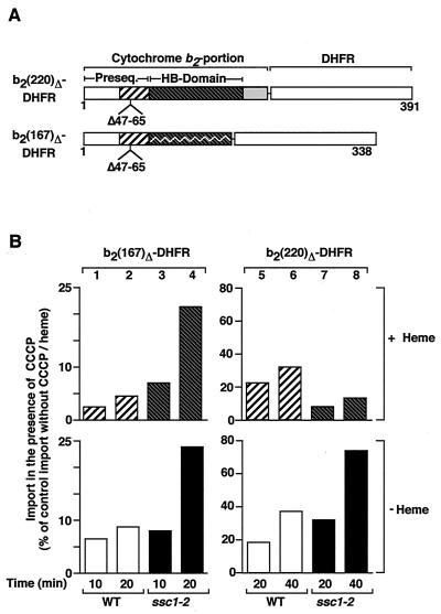
The mitochondrial heat shock protein Hsp70 (mtHsp70) is essential for driving translocation of preproteins into the matrix. Two models, trapping and pulling by mtHsp70, are discussed, but positive evidence for either model has not been found so far. We have analyzed a mutant mtHsp70, Ssc1-2, that shows a reduced interaction with the membrane anchor Tim44, but an enhanced trapping of preproteins. Unexpectedly, at a low inner membrane potential, ssc1-2 mitochondria imported loosely folded preproteins more efficiently than wild-type mitochondria. The import of a tightly folded preprotein, however, was not increased in ssc1-2 mitochondria. Thus, enhanced trapping by mtHsp70 stimulates the import of loosely folded preproteins and reduces the dependence on the import-driving activity of the membrane potential, directly demonstrating that trapping is one of the molecular mechanisms of mtHsp70 action.


Figures

FIG. 1
FIG. 1
Protein import into ssc1-2 mitochondria shows a lower sensitivity to a reduction of the inner membrane potential. (A) Isolated ssc1-2 and wild-type (WT) mitochondria were subjected to a heat shock at 37°C and subsequently incubated with reticulocyte lysate containing 35S-labeled b2(167)Δ-DHFR in the absence or presence of 15 μM CCCP at 25°C. The import reactions were stopped after the indicated times and treated with proteinase K to remove nonimported preproteins. After reisolation of the mitochondria, the import reactions were analyzed by SDS-PAGE and digital autoradiography. (B and C) b2(167)Δ-DHFR was imported into isolated mitochondria in the presence of the indicated concentrations of CCCP at 25°C for 5 min. The amount of imported protein was quantified by digital autoradiography. The amount of protein imported in the absence of CCCP was set to 100% (control). Bars indicate the standard errors of the means (from six independent experiments). (D) Calculation of the ratio of protein import into ssc1-2 versus wild-type mitochondria as quantified in panel C.
FIG. 2
FIG. 2
Enhancement of protein import in ssc1-2 mitochondria depends on the induction of the mtHsp70 mutant phenotype. (A) Comparison of the sensitivity to CCCP between wild-type and ssc1-2 mitochondria (Mitoch.). The membrane potential of isolated mitochondria from wild-type (WT) and ssc1-2 mitochondria (preincubated at 37°C) was assessed at 25°C in the absence (control) or presence of different concentrations of CCCP using the potential-dependent fluorescent dye DiSC3. The difference in fluorescence before and after the addition of valinomycin (Val.) represents an assessment of the magnitude of Δψ. To inhibit the F0F1-ATPase, oligomycin (20 μM) was included in the buffer. (B) Import stimulation depends on the induction of the mutant phenotype. Isolated wild-type and ssc1-2 mitochondria were heat-shocked for 15 min at 37°C prior to the import reaction or left at 25°C as indicated. b2(167)Δ-DHFR was imported into the mitochondria in the absence or presence of 15 μM CCCP. The import reactions were subsequently treated as described in the legend to Fig. 1. The ratio of protein import into ssc1-2 and wild-type mitochondria was determined.
FIG. 3
FIG. 3
Mutant mtHsp70 Ssc1-2 shows a differential interaction with the partner proteins Tim44, Mge1, and substrate proteins. (A) Interaction with Tim44 and Mge1. After incubation at 37°C, isolated wild-type (WT) and ssc1-2 mitochondria were lysed by Triton X-100 at the indicated concentrations of KCl and subjected to coimmunoprecipitation with antibodies directed against Tim44 (upper panel) or mtHsp70 (lower panel) as described in Materials and Methods. Upon SDS-PAGE, precipitated proteins were detected by Western blot analysis using antibodies against mtHsp70, Tim44, and Mge1, and 10% of the amount of lysed mitochondria is shown as a control. (B) Interaction with preprotein. The interaction of mtHsp70 with the preprotein b2(167)Δ-DHFR after import into wild-type and ssc1-2 mitochondria was assayed by coimmunoprecipitation with anti-mtHsp70 and digital autoradiography as described in Materials and Methods, and 2% of the total amount of the imported preprotein is shown as a control. p and i, precursor and intermediate forms of b2(167)Δ-DHFR, respectively.
FIG. 4
FIG. 4
Increased import of b2(167)Δ-DHFR into ssc1-2 mitochondria correlates with enhanced trapping by mtHsp70. (A and B) Imported b2(167)Δ-DHFR shows increased binding to the mutant mtHsp70 Ssc1-2. Radiolabeled b2(167)Δ-DHFR was imported into isolated mitochondria (after incubation at 37°C). The import was stopped after 5 min by addition of 1 μM valinomycin, and the mitochondria were further incubated at 25°C for the indicated times. The mitochondria were reisolated and lysed, and imported proteins bound to mtHsp70 were analyzed by coimmunoprecipitation, SDS-PAGE, and digital autoradiography. The amount of protein coprecipitated from wild-type (WT) mitochondria lysed directly after the addition of valinomycin (0 min) was set to 100% (control). i, matrix-targeted intermediate form. (C) The increased interaction of Ssc1-2 with imported preprotein is dependent on the induction of the temperature-sensitive phenotype. b2(167)Δ-DHFR was imported for 5 min into ssc1-2 and wild-type mitochondria that had been shifted to 37°C or kept at the permissive temperature prior to import. After the import reaction, the mitochondria were lysed and subjected to coimmunoprecipitation as described for panel A. The total amount of imported protein was set at 100%.
FIG. 5
FIG. 5
Preproteins in transit show an enhanced interaction with Ssc1-2. (A) Experimental approach. (B and C) b2(167)Δ-DHFR was imported for 5 min at 25°C into ssc1-2 and wild-type (WT) mitochondria (Mitoch.) in the presence of 5 μM MTX. Import was stopped by the addition of 1 μM valinomycin. One portion of the import reaction was treated with proteinase K (Prot. K in A, PK in B), and one was left untreated to assay the formation of membrane-spanning intermediates (B). The other portions of the import reaction were subjected to lysis and coimmunoprecipitation by antibodies directed against mtHsp70 either directly or after an additional incubation at 25°C (C). All samples were analyzed by SDS-PAGE and digital autoradiography. The total amount of accumulated protein was set at 100%.
FIG. 6
FIG. 6
Import of a tightly folded preprotein is not enhanced in ssc1-2 mitochondria. (A) Fusion proteins used in this experiment. b2(220)Δ-DHFR consists of residues 1 to 220 of the wild-type cytochrome b2 precursor fused to the entire mouse DHFR with a deletion of residues 47 to 65 in the presequence. The mature part (residues 81 to 220) contains a complete heme-binding (HB) domain (residues 81 to 181). b2(167)Δ-DHFR consists of residues 1 to 167 of cytochrome b2 (excluding residues 47 to 65) and DHFR. The truncation of the heme-binding domain leads to a less stable folding of this domain. (B) b2(167)Δ-DHFR and b2(220)Δ-DHFR were imported at 25°C into wild-type (WT) and ssc1-2 mitochondria under nonpermissive conditions in the presence or absence of 5 μM CCCP with or without the addition of 10 μM heme. The import reactions were subsequently treated as described in the legend to Fig. 1. The amount of protein imported into wild-type mitochondria in the absence of CCCP and heme after the maximal import time [20 min for b2(167)Δ-DHFR and 40 min for b2(220)Δ-DHFR] was set at 100% (control).

Similar articles

Cited by

References

    1. Alconada A, Gärtner F, Hönlinger A, Kübrich M, Pfanner N. Mitochondrial receptor complex from Neurospora crassa and Saccharomyces cerevisiae. Methods Enzymol. 1995;260:263–286. - PubMed
    1. Bauer M F, Hofmann S, Neupert W, Brunner M. Protein translocation into mitochondria: the role of TIM complexes. Trends Cell Biol. 2000;10:25–31. - PubMed
    1. Bauer M F, Sirrenberg C, Neupert W, Brunner M. Role of Tim23 as voltage sensor and presequence receptor in protein import into mitochondria. Cell. 1996;87:33–41. - PubMed
    1. Bömer U, Maarse A, Martin F, Geissler A, Merlin A, Schonfisch B, Meijer M, Pfanner N, Rassow J. Separation of structural and dynamic functions of the mitochondrial translocase: Tim44 is crucial for the inner membrane import sites in translocation of tightly folded domains, but not of loosely folded preproteins. EMBO J. 1998;17:4226–4237. - PMC - PubMed
    1. Bukau B, Horwich A. The Hsp70 and Hsp60 chaperone machines. Cell. 1998;92:351–366. - PubMed

Publication types

MeSH terms

LinkOut - more resources