. 2001 Sep;9(9):859-67.
doi: 10.1016/s0969-2126(01)00641-4.

The conformation of bound GMPPNP suggests a mechanism for gating the active site of the SRP GTPase

Affiliations

The conformation of bound GMPPNP suggests a mechanism for gating the active site of the SRP GTPase

S Padmanabhan et al. Structure. 2001 Sep.

Abstract

Background: The signal recognition particle (SRP) is a phylogenetically conserved ribonucleoprotein that mediates cotranslational targeting of secreted and membrane proteins to the membrane. Targeting is regulated by GTP binding and hydrolysis events that require direct interaction between structurally homologous "NG" GTPase domains of the SRP signal recognition subunit and its membrane-associated receptor, SR alpha. Structures of both the apo and GDP bound NG domains of the prokaryotic SRP54 homolog, Ffh, and the prokaryotic receptor homolog, FtsY, have been determined. The structural basis for the GTP-dependent interaction between the two proteins, however, remains unknown.

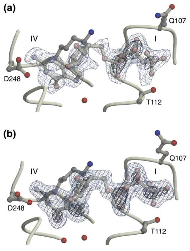
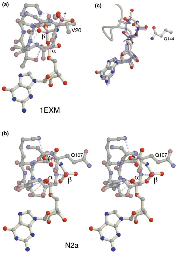
Results: We report here two structures of the NG GTPase of Ffh from Thermus aquaticus bound to the nonhydrolyzable GTP analog GMPPNP. Both structures reveal an unexpected binding mode in which the beta-phosphate is kinked away from the binding site and magnesium is not bound. Binding of the GTP analog in the canonical conformation found in other GTPase structures is precluded by constriction of the phosphate binding P loop. The structural difference between the Ffh complex and other GTPases suggests a specific conformational change that must accompany movement of the nucleotide from an "inactive" to an "active" binding mode.

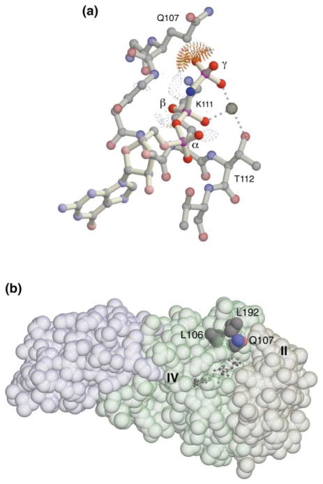
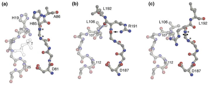
Conclusions: Conserved side chains of the GTPase sequence motifs unique to the SRP subfamily may function to gate formation of the active GTP bound conformation. Exposed hydrophobic residues provide an interaction surface that may allow regulation of the GTP binding conformation, and thus activation of the GTPase, during the association of SRP with its receptor.


Figures

Figure 1
Figure 1. GMPPNP Binding to the NG Domain
Omit difference (Fo − Fc) electron density maps contoured at 3 σ (light blue) and 6 σ (dark blue) for (a) structure N1 and (b) structure N2a. The triplet of electron-dense peaks to the right in each image indicates the positions of the phosphate groups. Two residues, Gln107 and Thr112, define the top and bottom of the P loop jaws.
Figure 2
Figure 2. Comparison of the Conformation of the GMPPNP with Other Structures
(a) The GMPPNP conformation in a complex with EF-Tu (1exm [7], representative of other GTPase structures). For clarity, only the side chains of Val20 (which corresponds in position to Gln107 of the SRP GTPase), Lys24, Thr 25, and Thr26 are shown. (b) A stereo image of the GMPPNP conformation observed in the NG complex. The orientation is approximately perpendicular to that in Figure 1. Only the side chains of Gln107, Lys111, Thr112, and Thr113 are shown. (c) Three structures (GDP, Mg2+GDP [2], and GMPPNP N2a) are shown superimposed after alignment of the core GTPase domain of each with the other. The two GDP conformations are shown in blue.
Figure 3
Figure 3. Steric Occlusion of the GTP Binding Site
(a) The Mg2+GMPPNP ligand complex modeled in the canonical conformation using the structure of the Mg2+GDP complex of NG as a starting point. Packing analysis carried out with Probe [17] illustrates the conflict that prevents movement of the γ-phosphate into the binding site—the spikes represent overlaps between the radii of the adjacent atoms (including hydrogens) [17]. The light dotted lens-shaped features generated by Probe indicate hydrogen bonds. (b) Conserved residues of the motif I P loop, Leu106, and Gln107, are interdigitated with Leu192 and the main chain backbone of motif III. The interaction constricts the P loop opening. The bound GMPPNP is indicated by the ball-and-stick model, and the positions of motifs IV and II that delimit the binding pocket are labeled. The three structural elements of the NG domain—the N domain helix bundle (left), the G domain, and the IBD insertion (right)—are indicated by distinct tints.
Figure 4
Figure 4. A Mechanism for Gating the NG Active Site
(a) The EF-Tu GMPPNP complex in the region corresponding to the SRP GTPase motifs I and III. The backbone and selected side chains (labeled in the figure) are shown. The backbone amide groups of motif III that play structurally and functionally similar roles in different GTPases are indicated with an asterisk. The positions of the β- and γ-phosphate groups in the EF-Tu structure are indicated. (b) The corresponding structure of GMPPNP complex N2a. Again, for clarity, only selected side chains (labeled in the figure) are shown. The hydrogen bond between the backbone amide of Gln107 and the carbonyl oxygen of Arg191 at the center of the interaction is indicated. The carbonyl oxygen of Gly190 (indicated by an arrow) is hydrogen bonded to the side chain of Arg191 [2]. The relative closing of the jaws of the P loop is apparent by comparison of the separation between residues 107 and 112 in (b) with that between the corresponding residues 20 and 25 in (a). (c) Structural consequences of a hypothesized 180° rotation of the ψ angle of Gly190. The motif III main chain takes on a conformation similar to that seen in other GTPases in the GTP bound state [14]. The conformation of the arginine side chain shown is speculative.

References

    1. Freymann DM, Keenan RJ, Stroud RM, Walter P. Structure of the conserved GTPase domain of the signal recognition particle. Nature. 1997;385:361–364. - PubMed
    1. Freymann DM, Keenan RJ, Stroud RM, Walter P. Functional changes in the structure of the SRP GTPase on binding GDP and Mg2+GDP. Nature Struct Biol. 1999;6:793–801. - PubMed
    1. Montoya G, Kaat KT, Moll R, Schäfer G, Sinning I. The crystal structure of the conserved GTPase of SRP54 from the archeon Acidianus ambivalens and its comparison with related structures suggests a model for the SRP-SRP receptor complex. Structure. 2000;8:515–525. - PubMed
    1. Montoya G, Svensson C, Luirink J, Sinning I. Crystal structure of the NG domain from the signal-recognition particle receptor FtsY. Nature. 1997;385:365–369. - PubMed
    1. Hirshberg M, Stockley RW, Dodson G, Webb MR. The crystal structure of human rac1, a member of the rho-family complexed with a GTP analogue. Nature Struct Biol. 1997;4:147–151. - PubMed

Publication types

MeSH terms

Substances