. 2001 Sep 17;20(18):5091-100.
doi: 10.1093/emboj/20.18.5091.

A split motor domain in a cytoplasmic dynein

Affiliations

A split motor domain in a cytoplasmic dynein

A Straube et al. EMBO J. .

Abstract

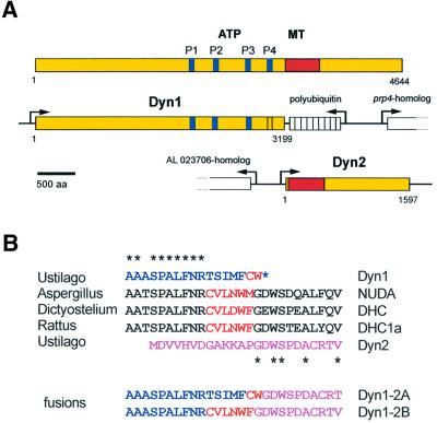
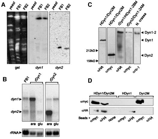
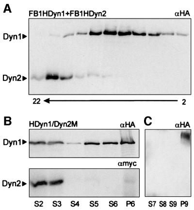
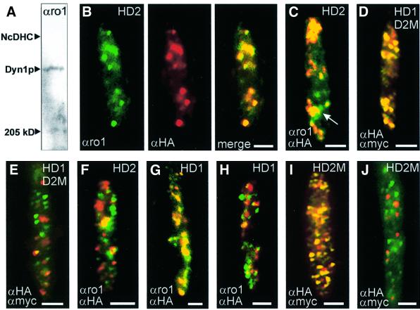
The heavy chain of dynein forms a globular motor domain that tightly couples the ATP-cleavage region and the microtubule-binding site to transform chemical energy into motion along the cytoskeleton. Here we show that, in the fungus Ustilago maydis, two genes, dyn1 and dyn2, encode the dynein heavy chain. The putative ATPase region is provided by dyn1, while dyn2 includes the predicted microtubule-binding site. Both genes are located on different chromosomes, are transcribed into independent mRNAs and are translated into separate polypeptides. Both Dyn1 and Dyn2 co-immunoprecipitated and co-localized within growing cells, and Dyn1-Dyn2 fusion proteins partially rescued mutant phenotypes, suggesting that both polypeptides interact to form a complex. In cell extracts the Dyn1-Dyn2 complex dissociated, and microtubule affinity purification indicated that Dyn1 or associated polypeptides bind microtubules independently of Dyn2. Both Dyn1 and Dyn2 were essential for cell survival, and conditional mutants revealed a common role in nuclear migration, cell morphogenesis and microtubule organization, indicating that the Dyn1-Dyn2 complex serves multiple cellular functions.


Figures

None
Fig. 1. Domain structure and organization of cytoplasmic dynein from U.maydis. (A) DHC from rat contains four P-loops (P1–4) that are also found in the predicted sequence of Dyn1, although the fourth P-loop differs from the consensus. Dyn1 lacks the putative MT-binding site (shown in red), which is encoded by a second gene, dyn2. (B) The transition from the C-terminus of Dyn1 (blue) to the N-terminus of Dyn2 (purple) lies within a highly conserved sequence region. Together, Dyn1 and Dyn2 contain most of the conserved amino acids (asterisks). However, typical cysteine and tryptophan residues are missing, or are misplaced at the C-terminus of Dyn1 (marked in red). The two fusion proteins generated in this study are either a direct fusion of Dyn1 and Dyn2, which eliminates the N-terminal 13 amino acids of Dyn2 (Dyn1-2A), or contain the missing amino acid stretch typical for cytoplasmic DHCs (Dyn1-2B).
None
Fig. 2. Southern, northern and western analysis of dynein from U.maydis. (A) Chromosomes were separated on a pulse field gel. After blotting, the same filter was incubated with a dyn1- and a dyn2-specific probe. Both genes hybridized to distinct chromosomes in U.maydis strains FB1 and FB2, but no signal was obtained with genomic DNA of Saccharomyces cerevisiae (yeast). (B) Northern blot of total RNA from wild type (FB1) and the conditional mutants in dyn1 (rDyn1) and dyn2 (rDyn2) was incubated simultaneously with 32P-labelled probes against dyn1 and dyn2. In FB1 two distinct mRNAs corresponding to the two genes are detected. In the conditional mutants at permissive conditions (ara) dyn1 and dyn2 are strongly over expressed. After 5 h in glucose-containing medium (glu) the amount of either dyn1 (rDyn1) or dyn2 (rDyn2) decreased significantly. (C) In western blots using epitope antibodies (αHA, αMyc) and extracts of strain FB2HDyn1/Dyn2M both polypeptides appear at their expected size of ∼360 kDa (HA-Dyn1) and ∼180 kDa (Dyn2-Myc), respectively. Both were significantly smaller than the DHC from N.crassa that was detected in extracts of N.crassa by an antibody against N.crassa dynein (αro1, kindly provided by M.Plamann). Integration of epitope-tagged Dyn1–Dyn2 fusion constructs in FB1rDyn1 (rDyn1/HDyn1-2BM) and FB1rDyn2 (rDyn2/Dyn1-2AM) led to the expression of large fusion proteins. (D) Extracts of the double-tagged strain FB2HDyn1/Dyn2M were incubated with magnetic beads loaded with antibodies directed against the Myc epitope (αMyc), the HA epitope (αHA) or no antibody (none). Each antibody precipitated both HA-Dyn1 and Dyn2-Myc. Neither antibodies nor beads non-specifically precipitated tagged Dyn1 or Dyn2.
None
Fig. 3. Dyn1 and Dyn2 behaviour in sucrose density gradients and MT affinity purification. (A) Extracts from FB1HDyn1 and FB1HDyn2 were loaded on 10–25% sucrose gradients and run for 13 h at 150 000 g. Fractions of 200 µl were collected and each second fraction was analysed by western analysis using αHA and αMyc antibodies. Note that HA-Dyn1 is completely separated from Dyn2-Myc, suggesting that the heavy chain complex dissociated during preparation. Fraction numbers are given below. (B) MT affinity experiments were performed using extracts of FB2HDyn1/Dyn2M. Both HA-Dyn1 and Dyn2-Myc can be detected in the same high speed supernatant (S2), but only a minor fraction binds to MTs and most protein remains in the following supernatant (S3). Washing the MT pellet released only small amounts of HA-Dyn1, while no Dyn2-Myc was found in S4. Surprisingly, treatment with 10 mM MgATP released Dyn1 (S5, S6), while only very small amounts of Dyn2 were present in the S5, S6 or final MT pellet (P6). (C) S5 and S6 were loaded on a 10–25% sucrose density gradient. After centrifugation the two peak fractions containing HA-Dyn1 were pooled and used for an additional round of MT binding. Again, HA-Dyn1 was able to bind MTs (P9) and no HA-Dyn1 remained in the supernatant (S7). However, HA-Dyn1 could not be released by MgATP (S8, S9).
None
Fig. 4. Localization of Dyn1 and Dyn2 in haploid U.maydis cells. (A) Western blot using the αro1 antibody that was raised against the P-loop region of the DHC from N.crassa. The antibody clearly recognized a band at the expected size for Dyn1. However, the signal on blots was weak and could not be used for further western analysis. The position of the DHC from N.crassa (NcDHC) and myosin (250 kDa) is indicated. (B) Co-localization of Dyn1 and HA-Dyn2 in yeast-like cells of strain FB2HDyn2. The αro1 antibody gave a punctuate staining of Dyn1 (green). The same distribution was seen for HA-Dyn2 visualized by αHA antibodies (red). The overlay revealed a clear co-localization of Dyn1 and Dyn2 (yellow). (C) Overlay of signals for Dyn1 and HA-Dyn2, detected by αro1 and αHA, in a growing cell of FB2HDyn2. At the onset of budding, signals for both polypeptides co-localize (yellow) and the complex is concentrated at the growth region of the cell. Occasionally, single spots of Dyn1 were detected (green, arrow). (D and E) Detection of HA-Dyn1 and Dyn2-Myc by specific antibodies directed against both epitope tags in cultures of FB2HDyn1/Dyn2M. In agreement with the results described above, signals for HA-Dyn1 and Dyn2-Myc co-localize (D). However, some unbudded cells showed a clear separation of most αHA (green dots) and αMyc signals (red dots), suggesting that the HA-Dyn1–Dyn2-Myc complex disassembled in these cells (E). (F) Localization of Dyn1 and HA-Dyn2 by αro1 and αHA in FB2HDyn2. Again, some unbudded cells showed a separation of Dyn1 (green dots) and HA-Dyn2 (red dots). (G and H) Detection of HA-Dyn1 by αro1 and αHA antibodies in strain FB1HDyn1. Most signals co-localize (G), however in some unbudded cells almost no correspondence was observed (H). (I and J) Detection of HA-Dyn2-Myc by αHA and αMyc antibodies in FB2HDyn2M. As expected, both antibodies recognize the double-tagged Dyn2 (I; overlay results in yellow). However, not all signals co-localize and in densely grown cultures no correspondence was seen (J). All bars correspond to 2 µm. Antibodies are given in the lower left, strains are indicated in the upper right (HD1, FB1HDyn1; HD2, FB2HDyn2; HD1D2M, FB2HDyn1/Dyn2M; HD2M, FB2HDyn2M).
None
Fig. 5. Nuclear migration in FB1rDyn2 expressing GFP fused to a nuclear localization signal. (A) Co-localization of nuclear DNA, stained with DAPI, and GFP fused to the GAL4 nuclear localization signal. The fusion protein localized to the nuclei of haploid U.maydis cells. (B) Nuclear distribution during the cell cycle of U.maydis. During polar budding the nucleus remains stationary in the middle of the mother cell, until pre-mitotic migration into the bud occurs. During mitosis the GFP signal disappears while a short spindle is formed (Steinberg et al., 2001; position of spindle is marked by an arrowhead). Finally, the two nuclei show post-mitotic migration and are positioned in the middle of mother and daughter cell, respectively. (C) Motion of tubular extensions at the nucleus. In growing cells, tubular extensions are pulled towards the bud (asterisk). Previous studies have indicated that the minus ends of MTs are located in the neck region (Steinberg et al., 2001), suggesting that a minus-end-directed motor is responsible for this motility. (D) Cells of strain FB1rDyn2/nGFP after 10 h in CM-G. All motion is gone and nuclei accumulate in the mother cell, indicating that dynein participates in nuclear migration. Bars in (A), (B) and (D) correspond to 5 µm, and in (C) to 2 µm.
None
Fig. 6. Nuclear distribution phenotype of conditional mutants in dyn1 and dyn2. (A) Wild-type strain FB1 as well as conditional mutant strains FB1rDyn1 and FB1rDyn2 show normal cell shape when grown under permissive conditions (CM-A). All strains contain a single nucleus positioned in the cell centre. After shift and growth under restrictive conditions (CM-G) expression of dyn1 and dyn2 is repressed, resulting in accumulations of nuclei in the mother cell due to a nuclear migration defect. Bars correspond to 5 µm. (B) Quantification of the nuclear migration defect in FB1rDyn1 and FB1rDyn2 after growth under restrictive conditions. The number of aberrant cells containing no, two or three and more nuclei began to increase after 8–10 h growth in CM-G. The graph is based on three experiments with 100 cells at each time point.
None
Fig. 7. Partial rescue of the morphological and nuclear phenotype of conditional Dyn2 by Dyn1–Dyn2 fusion proteins. (A) After 24 h in CM-G, cells of strain FB1rDyn2 change their morphology and tend to lose their cell polarity (arrow) while large bud-like extensions are formed (arrowhead). (B) Corresponding DAPI staining of DNA in FB1rDyn2. Note that numerous nuclei accumulate in the deformed mother cell. (C) In FB1rDyn2, nuclei tend to cluster within the cell, suggesting that no nuclear migration occurred. (D) Expression of the fusion protein HDyn1-2BM in strain FB1rDyn2/HDyn1-2BM leads to a partial rescue of morphological phenotype of FB1rDyn2, although cells grew slightly larger. (E and F) Corresponding DAPI staining of DNA revealed that most cells still contain numerous nuclei. However, nuclei are lined up in the presence of HDyn1-2BM and almost no clusters were observed (F). Bars in (A), (B), (D) and (E) correspond to 10 µm, and in (C) and (F) to 3 µm.
None
Fig. 8. Dyn1 and MTs in cells of wild-type FB1 and conditional mutant strain FB1rDyn2. (A) Triple staining of MTs, Dyn1 and nuclear DNA in FB1. During growth the MT cytoskeleton becomes polarized and a PTS appears at the growth region (green, arrow; Steinberg et al., 2001). Dyn1, detected with αro1 antibodies (red), co-localizes with these spherical structures (yellow). The nucleus, stained with DAPI, is shown in blue. (B) Double staining of MTs and Dyn1. At higher magnification, antibodies against tubulin (αtub, green) and DHC (αro1, red) clearly co-localize (yellow). (C) The MT cytoskeleton in an unbudded cell of FB1rDyn2 under permissive conditions. (D) Alterations of MT organization in FB1rDyn2 after 13 h in CM-G. Cells contain more and longer MTs. Occasionally, pairs of spherical tubulin structures that most likely represent MTOCs lose their polar localization (arrows). Bars in (A), (C) and (D) correspond to 3 µm, and in (B) to 1 µm.

Similar articles

Cited by

References

    1. Asai D.J and Koonce,M.P. (2001) The dynein heavy chain: structure, mechanics and evolution. Trends Cell Biol., 11, 196–202. - PubMed
    1. Banuett F. (1995) Genetics of Ustilago maydis, a fungal pathogen that induces tumors in maize. Annu. Rev. Genet., 29, 179–208. - PubMed
    1. Banuett F. and Herskowitz,I. (1989) Different a alleles of Ustilago maydis are necessary for maintenance of filamentous growth but not for meiosis. Proc. Natl Acad. Sci. USA, 86, 5878–5882. - PMC - PubMed
    1. Bölker M., Bohnert,H.U., Braun,K.H., Görl,J. and Kahmann,R. (1995) Tagging pathogenicity genes in Ustilago maydis by restriction-enzyme mediated integration (REMI). Mol. Gen. Genet., 248, 547–552. - PubMed
    1. Bottin A., Kämper,J. and Kahmann,R. (1996) Isolation of a carbon source-regulated gene from Ustilago maydis. Mol. Gen. Genet., 253, 342–352. - PubMed

Publication types

MeSH terms

Associated data