. 2001 Sep 17;20(18):5139-52.
doi: 10.1093/emboj/20.18.5139.

Net-targeted mutant mice develop a vascular phenotype and up-regulate egr-1

Affiliations

Net-targeted mutant mice develop a vascular phenotype and up-regulate egr-1

A Ayadi et al. EMBO J. .

Abstract

The ternary complex factors (TCFs) Net, Elk-1 and Sap-1 regulate immediate early genes through serum response elements (SREs) in vitro, but, surprisingly, their in vivo roles are unknown. Net is a repressor that is expressed in sites of vasculogenesis during mouse development. We have made gene-targeted mice that express a hypomorphic mutant of Net, Net delta, which lacks the Ets DNA-binding domain. Strikingly, homozygous mutant mice develop a vascular defect and up-regulate an immediate early gene implicated in vascular disease, egr-1. They die after birth due to respiratory failure, resulting from the accumulation of chyle in the thoracic cage (chylothorax). The mice have dilated lymphatic vessels (lymphangiectasis) as early as E16.5. Interestingly, they express more egr-1 in heart and pulmonary arteries at E18.5. Net negatively regulates the egr-1 promoter and binds specifically to SRE-5. Egr-1 has been associated with pathologies involving vascular stenosis (e.g. atherosclerosis), and here egr-1 dysfunction could possibly be associated with obstructions that ultimately affect the lymphatics. These results show that Net is involved in vascular biology and egr-1 regulation in vivo.


Figures

None
Fig. 1. Targeted mutagenesis of the net gene. (A) Schemes. Exon 2 contains the translation initiation codon and encodes amino acids 1–69 of Net. The 3′ probe for Southern blots, the 13 (wild-type) and 5 (mutant) kb XbaI fragments and the PCR primers (uc54, uc56 and uc57) are shown. B, BamHI; X, XbaI. (B) Southern analysis of XbaI-digested DNA from the progeny from a heterozygous (wild-type/mutant) inter-cross. The 3′ probe reveals 13 kb wild-type (WT) and 5 kb targeted (M) alleles. (C) PCR analysis of the same progeny. The products for the wild-type (WT, 1550 bp) and the targeted (M,1300 bp) alleles are indicated. (D) Analysis of net transcripts by RT–PCR. RNA from E16.5 wild-type and homozygous mutant embryos was used for RT–PCR with primers from exons 1–4. As expected, mutant RNA is not amplified with exon 2 primers due to the deletion, but is amplified with exon 3 + 4 primers. Exon 1 + 3 (ex1/ex3 set) primers produce a shorter product (90 bp) with the mutant as opposed to the wild-type (297 bp) RNA. The deduced mutant Net mRNA, Netδ, is shown. The in-frame ATG is indicated in italics. (E) Western blots of lung extracts from 2-week-old wild-type, heterozygous and homozygous mice. The #375 antibody, raised against a peptide encoded by exon 3 (Giovane et al., 1994, 1997), detects a 47 000 Da protein in wild-type (+/+) and heterozygous (+/δ) samples. The heterozygous (+/δ) and homozygous (δ/δ) samples have a 37 000 Da band, Netδ, as expected from initiation at the internal ATG. (F) Basal c-fos SRE reporter activity in transfected MEFs generated from wild-type and homozygous mutant (netδ/δ) embryos. The histogram shows the average absolute luciferase activity for three experiments repeated in triplicate.
None
Fig. 2. The phenotype of netδ/δ mice. (A) Genotype frequency of live-born animals from a heterozygous inter-cross. (B) Decreased survival of netδ/δ mice compared with +/+ and +/δ animals. (C) Typical phenotype developed by netδ/δ mice. This animal had respiratory distress at 6 days and died 2 days later. The thoracic cavity is full of chyle.
None
Fig. 3. Histology of the thorax. Haematoxylin–eosin-stained cross-sections of wild-type (A) and mutant (B) thoraxes are shown. Bars = 0.8 mm. The netδ/δ mouse was killed at 8 days, when it developed respiratory distress symptoms, together with a net+/+ littermate control. The pleural space is expanded in the mutant, filled by the chylous effusion (asterisk), which has compressed the lungs and the heart. (C and D) Magnifications of the dashed squares in (A) and (B), respectively. The netδ/δ thoracic wall (D) has dilated lymphatic vessels compared with the control (C). R, ribs; lv, lymphatic vessel. Bars = 0.2 mm.
None
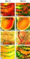
Fig. 4. Dilatation of the thoracic lymphatic vessels in netδ/δ mice. The LacZ-VEGFR-3 reporter strain was used to identify lymphatic vessels (Dumont et al., 1998). The lymphatic vessels of net+/+VEGFR-3+/– (A, C, E and G) and netδ/δVEGF-R3+/– mice (B, D, F and H) were stained with X-gal (blue). (B, D and F) Lymphatic staining of a netδ/δ mouse that developed chylothorax at 10 days of age. Lymphatics of the mutant thoracic wall (B) are dilated compared with the +/+ littermate (A). (C and D) Pericardial and (E and F) chest skin lymphatic vessels. Bars = 42 µm. (G and H) Thorax from a 5-day-old netδ/δ mouse before pleural effusion and a net+/+ littermate. R, ribs; ic, intercostal region. Bars = 90 µm.
None
Fig. 5. Co-expression of net and VEGFR-3 RNAs in E16.5 embryos. (A–F) ISH of sagittal sections with 35S-labelled riboprobes. The signal grains are white dots on a dark field. (A and D) Bright-field sections are shown for histology. (B and C) Net and the VEGFR-3 lymphatic marker are expressed in the thoracic duct (arrows). (E and F) Net is also expressed in lymphatic (arrows) and blood vessels in the gut. (G–I) Whole-mount ISH of thoracic biopsies. The specific signal has a dark blue colour. (G) Net is expressed in a plexus pattern (arrow). (H) The higher magnification shows Net staining of a lymphatic vessel (arrowhead). (I) Same magnification as (H), to show the aspect of lymphatics (arrowhead) in the thoracic wall stained with the VEGFR-3 probe. Ao, aorta; Oe, oesophagus; r, ribs; St, sternum; Td, thoracic duct; Tr, trachea; Ve, vertebrae. Bars = 50 µm (A–F), 200 µm (G), 100 µm (H and I).
None
Fig. 6. Unaltered expression of c-fos and egr-1 RNA in bony structures of netδ/δ E16.5 embryos. ISH of net+/+ (A, C, E, G and I) and netδ/δ (B, D, F, H and J) embryos. (C and E), (D and F), (I) and (J) are the dark fields corresponding to the bright fields (A), (B), (G) and (H), respectively. Expression of: (i) c-fos (C and D) and egr-1 (E and F) is not altered in the mutant embryo in the articular surface between the basioccipital bone and vertebrae (arrowheads); and (ii) of egr-1 (I and J) in the articular joint space from the hind limb (arrowheads). Ar, articular space; Bo, basioccipital bone; Br, brain; Hu, humerus; OC, otic capsule; Ra, radius; Ve, vertebrae. Bars = 90 µm (A–F) and 110 µm (G–J).
None
Fig. 7. Up-regulation of egr-1 RNA in the heart of netδ/δ E16.5 embryos. ISH on sagittal frozen sections of the thoracic region. (A and B) Bright fields, (C–H) corresponding dark fields for net+/+ (A, C, E and G) and netδ/δ (B, D, F and H) mice. (C and D) Egr-1 labelling is stronger in the atrial wall of the netδ/δ heart (arrowheads) but not, for example, in the ribs or the aorta. (E and F) VEGFR-1 endothelial marker. (G and H) Net expression. VEGFR-1 and net are expressed in the atrial wall (arrowheads, E–H). Ao, aorta; At, atrium; Ht, heart; Lu, lung; Ri, ribs; Th, thymus; Tr, trachea. Bar = 110 µm.
None
Fig. 8. Spatially restricted up-regulation of egr-1 RNA in the lung vasculature of netδ/δ E18.5 embryos. Egr-1 ISH on sagittal sections from the thoracic cage of net+/+ (B, E, H and K) and netδ/δ (C, F, I and L) embryos. The bright fields (A, D, G and J) of the net+/+ sections are shown for histology. Comparable sections (1–3) are shown for net+/+ and netδ/δ embryos. In section 1, egr-1 expression is similar in the wild-type and mutant mice, whereas in the other two sections there is stronger labelling in individual pulmonary arteries in the mutant (arrowheads; F and I). There is no difference in egr-1 labelling in other sites of expression, such as the thymus, the rib perichondrium and the walls of the vena cava as well as other vascular structures. (J, K and L) Higher magnification of sections 2, showing egr-1 up-regulation in the wall of an artery of a netδ/δ embryo (L, arrowheads), compared with the same vessel of a wild-type embryo (K, arrowheads), where egr-1 is detected within a restricted area. Ar, artery; Ht, heart; L, vascular lumen; Li, liver; Lu, lung; Ri, ribs; Th, thymus; VC, vena cava. Bars = 200 µm (A–I) and 25 µm (J–L).
None
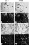
Fig. 9. Egr-1 ISH of lungs before the onset of chylothorax. Equivalent sagittal frozen sections from the thoracic region of 2-day-old net+/+ (A, C, E and G) and netδ/δ (B, D, F and H) mice. (C and D) Lower magnification showing a specific increase in the egr-1 signal throughout netδ/δ lungs (arrowheads). (E–H) Higher magnifications of similar regions of the lungs. (G and H) Patchy egr-1 up-regulation is detected in pulmonary blood vessels (arrowheads) and parenchyma (arrows). Ht, heart; Lu, lung; Th, thymus; Ri, ribs; VC, vena cava. Bars = 150 µm (A–D) and 50 µm (E–H).
None
Fig. 10. Net represses the activity of and binds to the egr-1 promoter. (A) Schemes of the mouse egr-1 promoter–luciferase reporters. Egr-1200-Luc contains 1200 bp of the egr-1 promoter with its five SREs (closed squares), whereas egr-250-Luc contains 250 bp upstream from the transcription start (arrows) with two proximal SREs. (B) Decreasing endogenous Net expression stimulates the egr-1 reporter through the distal SREs. NIH-3T3 cells were transfected in triplicate with egr-1 reporters and the p601D-anti-net plasmid that produces antisense net RNA, and luciferase activity was measured. An increasing amount of anti-net leads to significant and reproducible activation of the egr-1200-Luc reporter, but not egr-250-Luc. (C, D and E) Equal amounts of in vitro translated Net, Netδ and SRF proteins were used for gel retardation assays with wild-type or mutant SRE-5 probes (mut ets and mut SRF: mutated binding sites). Proteins and SRE-5 probes were incubated as indicated at the top and complexes were resolved by PAGE. Arrows indicate the complexes formed by Net, SRF and both proteins (TC, ternary complex), as well as a non-specific complex (NC) and the free probe (FP). (D) To compare off-rates, complexes were allowed to form for 25 min at 25°C and, after cooling on ice, a 500-fold excess of cold probe was added, the reactions were incubated for the indicated times at 0°C and then immediately run on the gel. 30′* was incubated for 30 min without competitor. (F) Nuclear extracts (Giovane et al., 1997) from net+/+ and netδ/δ lungs and in vitro translated Net and SRF proteins were used simultaneously for gel retardation assays with wild-type or mut ets SRE-5 probes. The Net antibodies were #375 (Giovane et al., 1994).

References

    1. al-Moslih M.I. and Dubes,G.R. (1973) The kinetics of DEAE–dextran-induced cell sensitization to transfection. J. Gen. Virol., 18, 189–193. - PubMed
    1. Arsenian S., Weinhold,B., Oelgeschlager,M., Ruther,U. and Nordheim,A. (1998) Serum response factor is essential for mesoderm formation during mouse embryogenesis. EMBO J., 17, 6289–6299. - PMC - PubMed
    1. Ayadi A., Suelves,M., Dolle,P. and Wasylyk,B. (2001) Net, an Ets ternary complex transcription factor, is expressed in sites of vasculogenesis, angiogenesis and chondrogenesis during mouse development. Mech. Dev., 102, 205–208. - PubMed
    1. Beddington R.S., Morgernstern,J., Land,H. and Hogan,A. (1989) An in situ transgenic enzyme marker for the midgestation mouse embryo and the visualization of inner cell mass clones during early organogenesis. Development, 106, 37–46. - PubMed
    1. Clarkson R.W., Shang,C.A., Levitt,L.K., Howard,T. and Waters,M.J. (1999) Ternary complex factors Elk-1 and Sap-1a mediate growth hormone-induced transcription of egr-1 (early growth response factor-1) in 3T3-F442A preadipocytes. Mol. Endocrinol., 13, 619–631. - PubMed

Publication types

MeSH terms