. 2001 Sep 17;20(18):5197-206.
doi: 10.1093/emboj/20.18.5197.

Independent function of two destruction domains in hypoxia-inducible factor-alpha chains activated by prolyl hydroxylation

Affiliations

Independent function of two destruction domains in hypoxia-inducible factor-alpha chains activated by prolyl hydroxylation

N Masson et al. EMBO J. .

Abstract

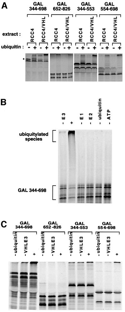
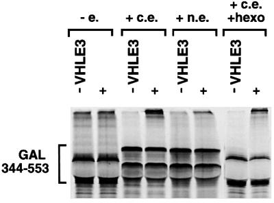
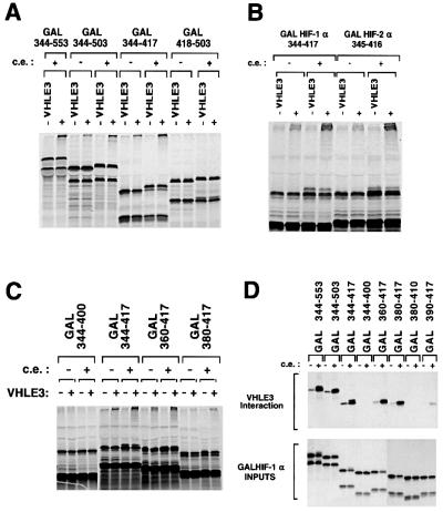
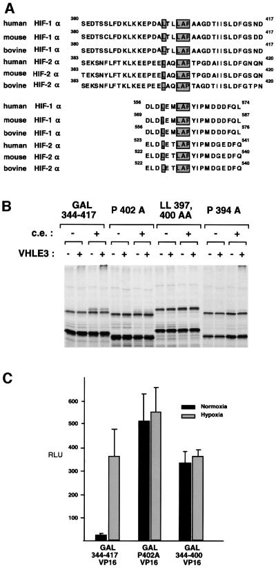
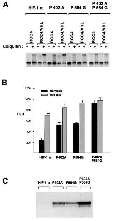
Oxygen-dependent proteolytic destruction of hypoxia-inducible factor-alpha (HIF-alpha) subunits plays a central role in regulating transcriptional responses to hypoxia. Recent studies have defined a key function for the von Hippel-Lindau tumour suppressor E3 ubiquitin ligase (VHLE3) in this process, and have defined an interaction with HIF-1 alpha that is regulated by prolyl hydroxylation. Here we show that two independent regions within the HIF-alpha oxygen-dependent degradation domain (ODDD) are targeted for ubiquitylation by VHLE3 in a manner dependent upon prolyl hydroxylation. In a series of in vitro and in vivo assays, we demonstrate the independent and non-redundant operation of each site in regulation of the HIF system. Both sites contain a common core motif, but differ both in overall sequence and in the conditions under which they bind to the VHLE3 ligase complex. The definition of two independent destruction domains implicates a more complex system of pVHL-HIF-alpha interactions, but reinforces the role of prolyl hydroxylation as an oxygen-dependent destruction signal.


Figures

None
Fig. 1. The VHLE3 ligase can interact functionally with two distinct regions of the HIF-1α ODDD. (A) Ubiquitylation of [35S]methionine-labelled GAL–HIF-1α fusion proteins by cytoplasmic extracts from VHL-defective RCC4 cells, or RCC4 cells stably transfected with pcDNA3-VHL (RCC4/VHL). Reactions were performed in the presence or absence of exogenous ubiquitin as indicated. VHLE3-dependent ubiquitylation, resulting in a strong signal of decreased mobility at the top of the lane, is seen when the substrate contained HIF-1α amino acids 344–698, 344–553 and 554–698, but not amino acids 652–826. The asterisk denotes the full-length, correctly initiated GAL344–698 fusion protein (see Materials and methods). (B) VHLE3-dependent ubiquitylation by purified components of the pathway. Ubiquitylation of an immunopurified GAL–HIF-1α fusion protein (GAL344–698) occurred in the presence of E1, E2, VHLE3 (E3), ubiquitin and ATP (lane marked ‘+’) but not in the absence of any individual component. (C) Ubiquitylation of different GAL–HIF-1α fusion proteins in the purified component assay. Reactions were performed using all components (+), or mixtures lacking either ubiquitin (–ubiquitin) or VHLE3 (–VHLE3). VHLE3-dependent ubiquitylation is seen when the substrate contained HIF-1α amino acids 344–698, 344–553 and 554–698, but not amino acids 652–826.
None
Fig. 2. Effect of pre-incubation of GAL344–553 substrate with cell extracts on VHLE3-dependent ubiquitylation. The substrate was incubated in buffer alone (–e.), or in cytoplasmic extract (+c.e.), nuclear extract (+n.e.) or cytoplasmic extract that had been depleted of ATP by incubation with hexokinase and glucose (+c.e. +hexo). All extracts were prepared from RCC4 cells lacking pVHL. Following immunopurification, the GAL344–553 substrates were ubiquitylated in the purified component assay in the presence (+) or absence (–VHLE3) of VHLE3. Pre-treatment with cytoplasmic extract greatly enhanced VHLE3-dependent ubiquitylation.
None
Fig. 3. Determination of HIF-α sequences that support VHLE3 interaction and ubiquitylation. (A–C) Ubiquitylation of GAL-HIF-1α or GAL–HIF-2α fusion protein substrates that had been pre-incubated in buffer alone (–c.e.) or in cytoplasmic extract (+c.e.), immunopurified and then added to reaction mixes either containing (+) or lacking VHLE3 (–VHLE3). (A) Exon-based analysis demonstrating that sequences encoded by HIF-1α exon 9 (residues 344–417) constitute an N-terminal ODDD target site for VHLE3-dependent ubiquitylation. Note that the actual exon 10–11 boundary is at amino acids 512–513. (B) Assay of homologous HIF-1α and HIF-2α sequences indicating that this VHLE3 target site is conserved between the HIF-α isoforms. (C) Further analysis of the N-terminal ODDD target site indicating that HIF-1α residues 360–417 are required for efficient ubiquitylation, whereas residues 380–417 support reduced but still significant VHLE3-dependent ubiquitylation. (D) VHLE3 interaction assay. The indicated [35S]methionine-labelled GAL–HIF-1α fusion proteins were incubated either in buffer alone (–c.e.) or in cytoplasmic extract (+c.e.). 786-0 HA⋅VHL cell extract (prepared in buffer that does not support VHLE3 target site modification) was then added and anti-HA immunoprecipitation performed. The retrieved immunoprecipitates and input samples of the GAL–HIF-1α fusion proteins were analysed by SDS–PAGE and autoradiography. HIF-1α residues 380–417 constitute a minimal domain capable of interaction with VHLE3 after exposure to c.e.
None
Fig. 4. Identification of a potential core motif targeted by VHLE3. (A) The amino acid sequences of the N- and C-terminal VHLE3-binding sites identified in human HIF-1α are aligned with the corresponding regions in other species (as indicated) and with HIF-2α sequences. A core motif is shaded. (B) Effect of mutations in this core motif on VHLE3-dependent ubiquitylation. The GAL344–417 substrate and the indicated mutant derivatives were pre-treated with buffer alone (–) or with cytoplasmic extract (+), immunopurified then added to ubiquitylation reactions that did (+) or did not (–) contain VHLE3. Mutants P402A and LL397,400AA, but not P394A, ablate activity. (C) Oxygen-regulated activity of GAL–HIF-1α–VP16 fusion proteins containing the indicated HIF-1α sequences, in U2OS cells co-transfected with the GAL reporter pUAS-tk-Luc. Columns show corrected luciferase activity, mean ± 1 SE of three independent experiments. Regulated fusion protein activity is observed with HIF-1α residues 344–417, but not a P402A mutant derivative, or HIF-1α residues 344–400.
None
Fig. 5. Proline residues 402 and 564 are critical for HIF-1α regulation both in vitro and in vivo. (A) Ubiquitylation of wild-type HIF-1α or the indicated mutants by cytoplasmic extracts from RCC4 or RCC4/VHL cells in the presence (+) or absence (–) of exogenous ubiquitin. The double mutant P402A + P564G shows no VHLE3-dependent ubiquitylation, but single mutations of the critical proline residues at each individual VHLE3 target site only partially reduce ubiquitylation. (B) Activity of transiently transfected wild-type HIF-1α and the indicated mutant derivatives, in HIF-1α-deficient CHO-Ka13 cells co-transfected with the HRE reporter plasmid pGL3PGK6TKp. Columns show corrected luciferase activity, mean ± 1 SE of three independent experiments. The single proline mutants show partially enhanced normoxic activity compared with the wild-type HIF-1α, whereas the combined mutant showed full constitutive activity in normoxia. (C) Immunoblot analysis of transiently transfected wild-type HIF-1α and the indicated mutant derivatives, in extracts from HIF-1α-deficient CHO-Ka13 cells grown under normoxic conditions.
None
Fig. 6. The N-terminal VHLE3 target site is regulated by prolyl hydroxylation. (A) Effect of iron (II) supplementation on VHLE3-dependent ubiquitylation. GAL344–417 and GAL554–698 substrates were pre-incubated in buffer alone (–), in cytoplasmic extract (+) or in cytoplasmic extract with added iron (Fe2++). Following immunopurification, substrates were ubiquitylated in the purified component assay in the presence (+) or absence (–) of VHLE3. Both substrates exhibit VHLE3-dependent ubiquitylation, which is enhanced by pre-treatment with additional iron. (B) Effect of the proline analogue 3,4-dehydro-l-proline on VHLE3 interaction. [35S]methionine-labelled GAL–HIF-1α substrates were prepared in reactions containing excess l-proline (P) or 3,4-dehydro-l-proline (D), and tested for VHLE3 interaction using 786-0 HA⋅VHL cell extract as in Figure 3D (see Materials and methods). Autoradiographs show SDS–PAGE analyses of the interacting GAL–HIF-1α proteins in anti-HA immunoprecipitates, together with the input samples. (C) Effect of the prolyl hydroxylase inhibitor N-oxalylglycine on modification of GAL380–417 P394A substrate. N-oxalylglycine (1 mM) inhibited the modifying activity of cytoplasmic extract and prevented VHLE3 interaction. Modification was partially restored by the addition of 5 mM 2-oxoglutarate. (D) Interaction of VHLE3 with synthetic peptides. Biotinylated peptides corresponding to HIF-1α residues 390–417 (B28PRO), or the corresponding peptide with Pro402 substituted with hydroxyproline (B28HYP), were incubated with buffer alone (–c.e.) prior to incubation with 786-0 HA⋅VHL cell extract. Following retrieval of peptide using streptavidin beads, captured HA⋅VHL was detected by anti-HA immunoblotting. B28HYP but not B28PRO captured HA⋅VHL. B28PRO was also incubated with cytoplasmic extract (+c.e.) prior to VHLE3 interaction. (E) Excess B28HYP but not B28PRO blocks VHLE3-dependent ubiquitylation. GAL344–553 substrate was pre-treated with buffer alone (–c.e.) or with cytoplasmic extract (+c.e.), then added to the purified component ubiquitylation assay in the presence or absence of peptide as indicated.
None
Fig. 7. Different requirements for targeting of N- and C-terminal sites in the HIF-1α ODDD by VHLE3. (A) Comparison of unfractionated and S100 cytoplasmic extract. [35S]methionine-labelled GAL344–553 and GAL554–698 substrates were ubiquitylated in fresh cytoplasmic extract (+), cytoplasmic extract that had been left at 4°C for 4 h (4°C 4 h+) or the S100 supernatant of cytoplasmic extract (S100+) from RCC4 or RCC4/VHL cells. The S100 extract supported VHLE3-dependent ubiquitylation of GAL554–698 but not GAL344–553. (B) Interaction of GAL-HIF–1α with HA⋅VHL produced in reticulocyte lysate. [35S]methionine-labelled C-terminal HA-tagged VHL (VHLHA) or N-terminal HA-tagged VHL (data not shown) and non-radiolabelled GAL–HIF-1α substrates were mixed and incubated either in buffer (–) or in cytoplasmic extract (+) prior to immunoprecipitation using anti-GAL. Co-immunoprecipitation of VHLHA occurred with GAL554–698, whilst background levels of binding were obtained with GAL652–826 and GAL344–553. Equivalent immunoprecipitation of the GAL–HIF-1α proteins was confirmed by anti-GAL immunoblotting (data not shown).

References

    1. Berra E., Milanini,J., Richard,D.E., LeGall,M., Vinals,F., Gothie,E., Roux,D., Pages,G. and Pouyssegur,J. (2000) Signaling angiogenesis via p42/p44 MAP kinase and hypoxia. Biochem. Pharmacol., 60, 1171–1178. - PubMed
    1. Bhattacharya S., Michels,C.L., Leung,M.-K., Arany,Z.P., Kung,A.L. and Livingston,D.M. (1999) Functional role of p35srj, a novel p300/CBP binding protein, during transactivation by HIF-1. Genes Dev., 13, 64–75. - PMC - PubMed
    1. Cockman M.E. et al. (2000) Hypoxia inducible factor-α binding and ubiquitylation by the von Hippel–Lindau tumor suppressor protein. J. Biol. Chem., 275, 25733–25741. - PubMed
    1. Ema M., Hirota,K., Mimura,J., Abe,H., Yodoi,J., Sogawa,K., Poellinger,L. and Fujii-Kuriyama,Y. (1999) Molecular mechanisms of transcription activation by HLF and HIF1α in response to hypoxia: their stabilization and redox signal-induced interaction with CBP/p300. EMBO J., 18, 1905–1914. - PMC - PubMed
    1. Huang L.E., Arany,Z., Livingston,D.M. and Bunn,H.F. (1996) Activation of hypoxia-inducible transcription factor depends primarily on redox-sensitive stabilization of its α subunit. J. Biol. Chem., 271, 32253–32259. - PubMed

Publication types

MeSH terms