. 2001 Sep 17;20(18):5207-18.
doi: 10.1093/emboj/20.18.5207.

Structure of the yeast nucleosome core particle reveals fundamental changes in internucleosome interactions

Affiliations

Structure of the yeast nucleosome core particle reveals fundamental changes in internucleosome interactions

C L White et al. EMBO J. .

Abstract

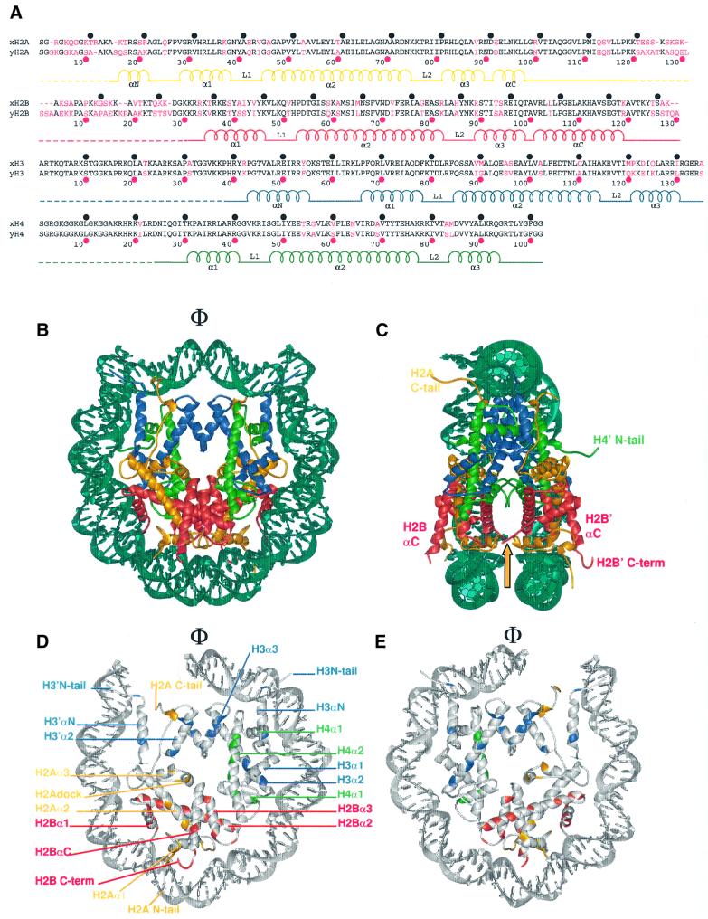
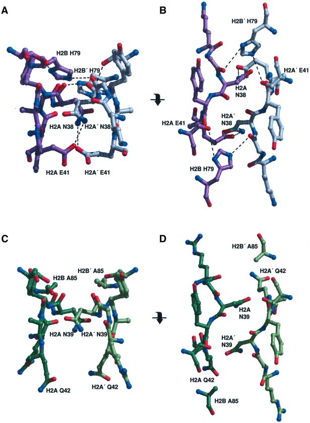
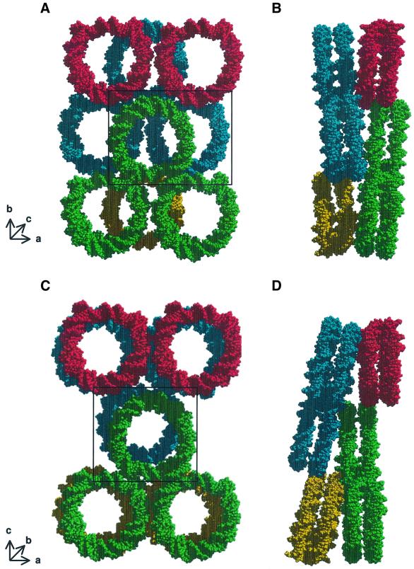
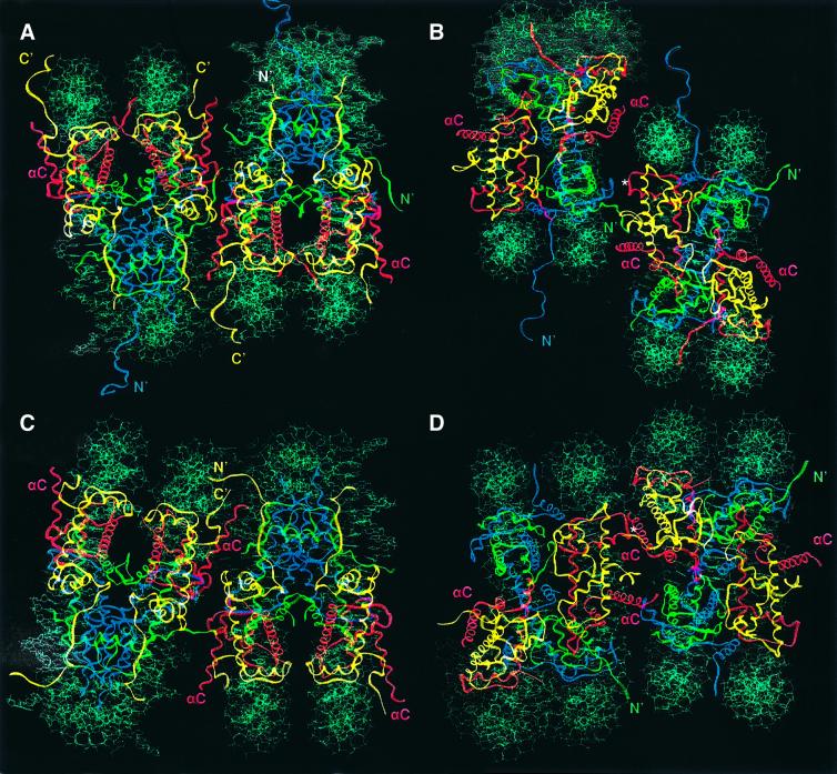
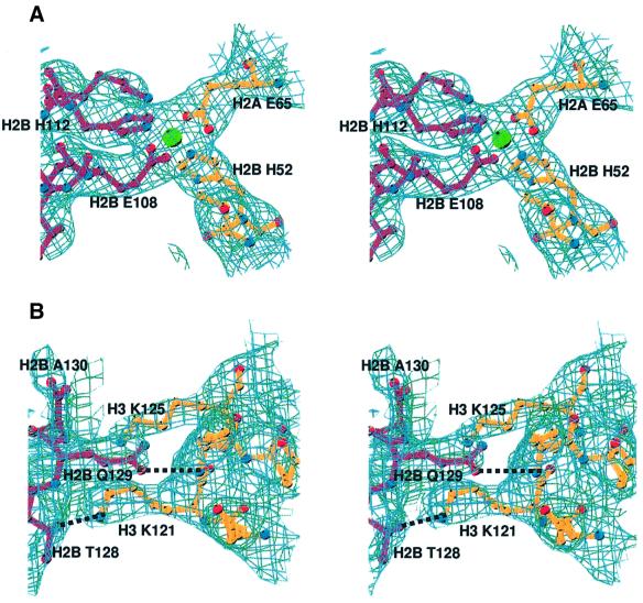
Chromatin is composed of nucleosomes, the universally repeating protein-DNA complex in eukaryotic cells. The crystal structure of the nucleosome core particle from Saccharomyces cerevisiae reveals that the structure and function of this fundamental complex is conserved between single-cell organisms and metazoans. Our results show that yeast nucleosomes are likely to be subtly destabilized as compared with nucleosomes from higher eukaryotes, consistent with the idea that much of the yeast genome remains constitutively open during much of its life cycle. Importantly, minor sequence variations lead to dramatic changes in the way in which nucleosomes pack against each other within the crystal lattice. This has important implications for our understanding of the formation of higher order chromatin structure and its modulation by post-translational modifications. Finally, the yeast nucleosome core particle provides a structural context by which to interpret genetic data obtained from yeast. Coordinates have been deposited with the Protein Data Bank under accession number 1ID3.


Figures

None
Fig. 1. Secondary and tertiary structure of the yeast nucleosome core particle. (A) Sequence alignment of X.laevis (top line) and S.cerevisiae histone proteins (bottom line). Amino acid differences are colored in magenta. Intervals of 10 amino acids for X.laevis (black circles) and S.cerevisiae (magenta circles) are indicated. The α-helices and loops located within the structured regions are labeled, and the flexible histone tails are indicated by dashed lines. (B) The crystal structure of the yeast nucleosome core particle, viewed down the superhelical axis. Histone chains are colored yellow for H2A, red for H2B, blue for H3 and green for H4. The DNA is shown in turquoise. α-helices and the location of the N- and C-terminal tails are shown. The position of the molecular dyad axis is indicated (Φ). (C) Side view of the yeast nucleosome core particle, obtained by rotation of 90° around the axis of non-crystallographic symmetry, with part of the DNA removed for clarity. The arrow denotes the location of the L1 loop. (D and E) Amino acid differences in the yeast octamer [as shown in (A)] are colored according to the histone coloring scheme in (B). The conserved amino acids and DNA are shown in gray. Only 73 bp of the DNA and associated proteins are shown. (D) The solvent-exposed surface view of one half of the nucleosome is shown, while (E) shows the same half of the nucleosome viewed from the interior surface between the two gyres of the DNA supercoil.
None
Fig. 2. Interactions between two H2A–H2B dimers within a nucleosome. (A) The side chain interactions of the H2A and H2A′ L1 loops in Xla-NCP, in the same orientation as shown in Figure 1C. Xla-H2A and H2A′ are colored in purple and gray, respectively. Dashed lines indicate hydrogen bonds. (B) The same region is shown in a view obtained by a 90° rotation around the horizontal axis (as indicated). (C and D) The equivalent region in Sce-NCP, viewed in the same orientation as in (A) and (B), respectively. Note the total absence of intermolecular hydrogen bonds.
None
Fig. 3. Crystal packing of X.laevis and S.cerevisiae nucleosome core particles. (AXla-NCP crystal packing viewed approximately down the superhelical axis. Short arrows show the approximate location of the three crystallographic axes. Only the DNA is shown for clarity; the same colors denote nucleosomes that lie within the same plane. (B) The same arrangement of molecules is rotated by 90° around the crystallographic b-axis. (C and DSce-NCP crystal packing in the same views as (A) and (B), respectively. The discrepancy in the notation of the crystallographic b- and c-axes stems from the fact that the previous study used different programs to index the data (Luger et al., 1997a). Particles whose molecular interaction is shown in Figure 4 are boxed.
None
Fig. 4. Protein–protein interactions within the crystal lattice. (A) Crystal contacts between two neighboring nucleosomes in X.laevis in a view that places the superhelical axis in a horizontal orientation (as seen in Figure 1C). (B) The same Xla-NCP packing as shown in (A), but viewed down the molecular 2-fold axis. This view is achieved by a 90° rotation around the superhelical axis (horizontal). In both views, the nucleosome core particle to the left of the pair corresponds to the center green particle in Figure 3A, whereas the right-hand particle corresponds to the blue particle in Figure 3A (boxed in Figure 3A and C, respectively). Histones are colored as in Figure 1. The location of the H2A and H4 histone tails is indicated. The position of the Mn2+ ion that is crucially involved in forming crystal contacts is shown (*). (C and D) Two yeast nucleosome core particles shown in the same orientation as seen in (A) and (B), respectively. With respect to Figure 3C, the same two particles are depicted as for Xla-NCP.
None
Fig. 5. Details of nucleosome–nucleosome interactions in yeast NCP. (A) Stereo view of a section of the |2FoFc| electron density map, calculated at 3.1 Å and contoured at 1σ, showing an Mn2+-mediated crystal contact between H2A Glu65 and H2B His52 of one nucleosome (dark red) and H2B′ Glu108 and H2B′ His112 of the neighboring nucleosome (gold). (B) Stereo view of the hydrogen bonds between the H2B C-terminal end of H2B αC (dark red) and residues in histone H3 of a neighboring nucleosome core particle (gold), showing a section of the |2FoFc| electron density map calculated at 3.1 Å and contoured at 8–1σ.

References

    1. Aalfs J.D. and Kingston,R.E. (2000) What does ‘chromatin remodeling’ mean? Trends Biochem. Sci., 25, 548–555. - PubMed
    1. Baer B.W. and Rhodes,D. (1983) Eukaryotic RNA polymerase II binds to nucleosome cores from transcribed genes. Nature, 301, 482–488. - PubMed
    1. Baxevanis A.D. and Landsman,D. (1998) Histone Sequence Database: new histone fold family members. Nucleic Acids Res., 26, 372–375. - PMC - PubMed
    1. Baxevanis A.D., Arents,G., Moudrianakis,E.N. and Landsman,D. (1995) A variety of DNA-binding and multimeric proteins contain the histone fold motif. Nucleic Acids Res., 23, 2685–2691. - PMC - PubMed
    1. Brünger A.T., Adams,P.D. and Rice,L.M. (1997) New applications of simulated annealing in X-ray crystallography and solution NMR. Structure, 5, 325–336. - PubMed

Publication types

Associated data