. 2001 Oct;183(20):5834-9.
doi: 10.1128/JB.183.20.5834-5839.2001.

Measuring virulence factor expression by the pathogenic bacterium Photorhabdus luminescens in culture and during insect infection

Affiliations

Measuring virulence factor expression by the pathogenic bacterium Photorhabdus luminescens in culture and during insect infection

P J Daborn et al. J Bacteriol. 2001 Oct.

Abstract

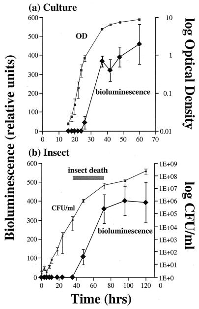
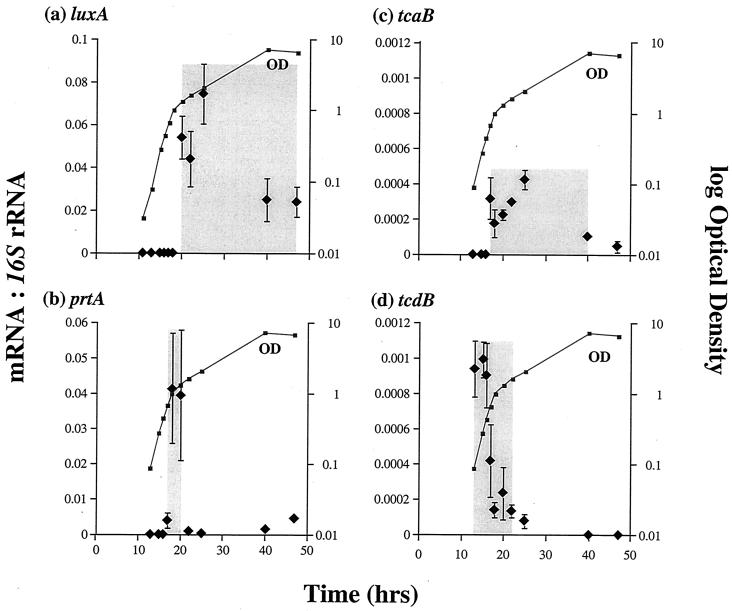
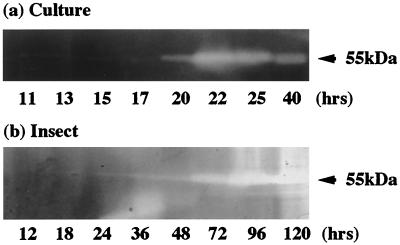
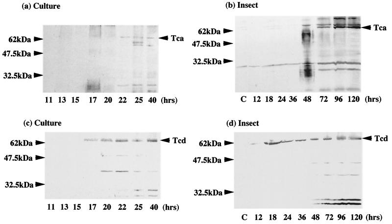
During insect infection Photorhabdus luminescens emits light and expresses virulence factors, including insecticidal toxin complexes (Tcs) and an RTX-like metalloprotease (Prt). Using quantitative PCR and protein assays, we describe the expression patterns of these factors both in culture and during insect infection and compare them to the associated bacterial growth curves. In culture, light and active Prt protease are produced in stationary phase. Tca also appears in stationary phase, whereas Tcd is expressed earlier. These patterns seen in a culture flask are strikingly similar to those observed during insect infection. Thus, in an infected insect, bacteria grow exponentially until the time of insect death at approximately 48 h, when both light and the virulence factors Prt protease and Tca are produced. In contrast, Tcd appears much earlier in insect infection. However, at present, the biological significance of this difference in timing of the production of the two toxins in unclear. This is the first documentation of the expression of Tcs and Prt in an insect and highlights the malleability of Photorhabdus as a model system for bacterial infection.


Figures

FIG. 1
FIG. 1
Growth curves for P. luminescens in culture and during an insect infection. (a) Growth curve in culture derived from measurements of OD over time and the associated bioluminescence produced. Note that light production peaks as the cells enter stationary phase. (b) Bacterial growth curve during an insect infection, derived from the mean number of CFU recovered from infected insects at different time points after injection of bacteria. The range of time (shaded bar) during which the study insects died is shown (actual mortality at 36, 48, and 72 h was 4, 60, and 100%, respectively). Note that light production peaks as the bacterial growth rate decreases. All data are shown as the means of three experiments with associated standard errors.
FIG. 2
FIG. 2
Relative abundance and times of expression (shaded areas) of luxA (a), prtA (b), tcaB (c), and tcdB (d) mRNAs versus 16S rRNA over time in culture. Note that tcdB message is transcribed at an earlier stage in culture growth than that of tcaB (see the text for discussion). All data are shown as the means of three experiments with their associated standard errors.
FIG. 3
FIG. 3
Detection by zymogram of the active Prt protease in culture supernatants and during an insect infection. Note the zone of protein clearing at ≈55 kDa, corresponding to production of the active Prt protease (see the text for discussion). Note also that the time scales differ for the culture experiment and the insect infection.
FIG. 4
FIG. 4
Western blot analysis of culture (a) and infected insects (b) with anti-Tca antibody and culture (c) and infected insects (d) with anti-Tcd antibody. Note that species cross-reactive with the anti-Tcd antibody can be detected after 17 h in culture, correlating with the peak of tcd transcription (Fig. 2d). Similarly, in an infected insect, Tcd can be detected after 18 h from the initial infection, before the time of insect death. Tca, in contrast, appears later (72 h postinfection) and in greater relative abundance when all the insects are dead.

References

    1. Bowen D, Blackburn M, Rocheleau T, Grutzmacher C, ffrench-Constant R H. Secreted proteases from Photorhabdus luminescens: separation of the extracellular proteases from the insecticidal Tc toxin complexes. Insect Biochem Mol Biol. 2000;30:69–74. - PubMed
    1. Bowen D, Rocheleau T A, Blackburn M, Andreev O, Golubeva E, Bhartia R, ffrench-Constant R H. Insecticidal toxins from the bacterium Photorhabdus luminescens. Science. 1998;280:2129–2132. - PubMed
    1. Bowen D J, Ensign J C. Purification and characterization of a high-molecular-weight insecticidal protein complex produced by the entomopathogenic bacterium Photorhabdus luminescens. Appl Environ Microbiol. 1998;64:3029–3035. - PMC - PubMed
    1. Colepicolo P, Cho K W, Poinar G O, Hastings J W. Growth and luminescence of the bacterium Xenorhabdus luminescens from a human wound. Appl Environ Microbiol. 1989;55:2601–2606. - PMC - PubMed
    1. Ehlers R U, Niemann I. Molecular identification of Photorhabdus luminescens strains by amplification of specific fragments of the 16S ribosomal DNA. Syst Appl Microbiol. 1998;21:509–519. - PubMed

Publication types

LinkOut - more resources