. 2001 Oct;183(20):5927-36.
doi: 10.1128/JB.183.20.5927-5936.2001.

Nonspecific adherence and fibril biogenesis by Actinobacillus actinomycetemcomitans: TadA protein is an ATPase

Affiliations

Nonspecific adherence and fibril biogenesis by Actinobacillus actinomycetemcomitans: TadA protein is an ATPase

M K Bhattacharjee et al. J Bacteriol. 2001 Oct.

Abstract

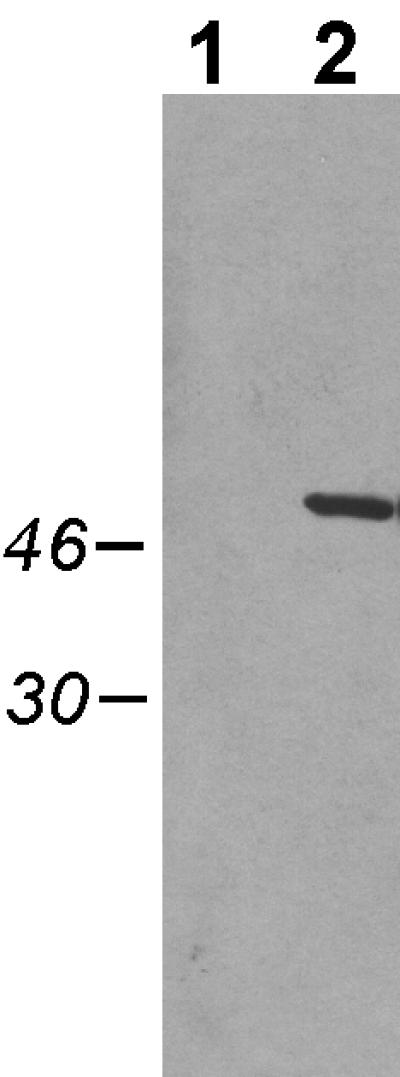
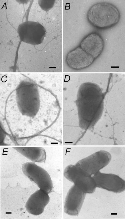
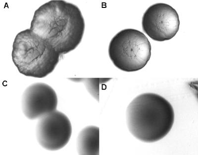
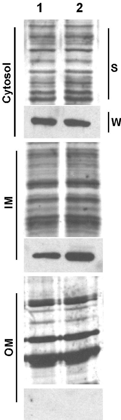
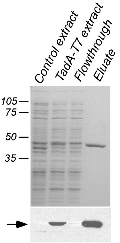
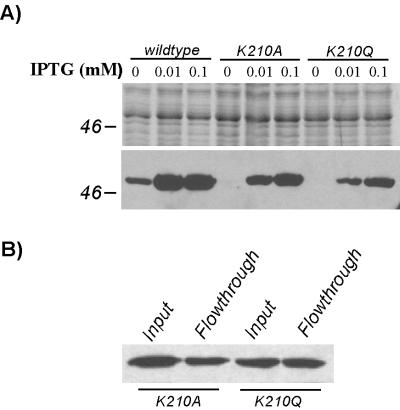
Cells of Actinobacillus actinomycetemcomitans, a gram-negative pathogen responsible for an aggressive form of juvenile periodontitis, form tenaciously adherent biofilms on solid surfaces. The bacteria produce long fibrils of bundled pili, which are required for adherence. Mutations in flp-1, which encodes the major subunit of the pili, or any of seven downstream tad genes (tadABCDEFG) cause defects in fibril production, autoaggregation, and tenacious adherence. We proposed that the tad genes specify part of a novel secretion system for the assembly and transport of Flp pili. The predicted amino acid sequence of TadA (426 amino acids, 47,140 Da) contains motifs for nucleotide binding and hydrolysis common among secretion NTP hydrolase (NTPase) proteins. In addition, the tadA gene is the first representative of a distinct subfamily of potential type IV secretion NTPase genes. Here we report studies on the function of TadA. The tadA gene was altered to express a modified version of TadA that has the 11-residue epitope (T7-TAG) fused to its C terminus. The TadA-T7 protein was indistinguishable from the wild type in its ability to complement the fibril and adherence defects of A. actinomycetemcomitans tadA mutants. Although TadA is not predicted to have a transmembrane domain, the protein was localized to the inner membrane and cytoplasmic fractions of A. actinomycetemcomitans cells, indicating a possible peripheral association with the inner membrane. TadA-T7 was purified and found to hydrolyze ATP in vitro. The ATPase activity is stimulated by Triton X-100, with maximal stimulation at the critical micellar concentration. TadA-T7 forms multimers that are stable during sodium dodecyl sulfate-polyacrylamide gel electrophoresis in nonreducing conditions, and electron microscopy revealed that TadA-T7 can form structures closely resembling the hexameric rings of other type IV secretion NTPases. Site-directed mutagenesis was used to substitute Ala and Gln residues for the conserved Lys residue of the Walker A box for nucleotide binding. Both mutants were found to be defective in their ability to complement tadA mutants. We suggest that the ATPase activity of TadA is required to energize the assembly or secretion of Flp pili for tight adherence of A. actinomycetemcomitans.


Figures

FIG. 1
FIG. 1
Expression of TadA-T7 in A. actinomycetemcomitans visualized by Western blot analysis. Cells were grown overnight in the presence of 0.1 mM IPTG, pelleted, and then resuspended in SDS loading dye, boiled, and separated by SDS-PAGE through a 12% polyacrylamide gel, as described in Materials and Methods. Lane 1, strain CU1000N carrying pJAK16 (vector); lane 2, strain CU1000N carrying pSK174 (tadA-T7). Numbers (left), molecular masses of relevant markers in kilodaltons.
FIG. 2
FIG. 2
Complementation of a tadA mutation for fibril formation. Cells were stained with 1% uranyl acetate and then viewed by electron microscopy, as described in Materials and Methods. (A) CU1000N tadA+; (B to F) AA1360 mutant with pJAK16 vector (B), wild-type tadA plasmid pEK2 (C), wild-type tadA-T7 plasmid pSK174 (D), tadA-T7(K210A) plasmid pSK199 (E), and tadA-T7(K210Q) plasmid pSK194 (F). Scale bars, 200 nm.
FIG. 3
FIG. 3
Complementation of the tadA mutation with tadA-T7 for rough-colony morphology. (A) CU1000N tadA+; (B to D) AA1360 tadA mutant with wild-type tadA-T7 plasmid pSK174 (B), tadA-T7(K210A) plasmid pSK199 (C), and tadA-T7(K210Q) plasmid pSK194 (D). The rough appearance of the colonies in panel B is typical for complemented tad mutations, as previously described (31).
FIG. 4
FIG. 4
Cellular location of TadA-T7. Fractionation of cells into cytoplasmic (cytosol), inner membrane (IM), and outer membrane (OM) fractions was done by the method of Haase et al. (21), as described in Materials and Methods. All strains carry the TadA-T7-expressing plasmid. Samples were separated by SDS-PAGE through a 12% polyacrylamide gel. Lane 1, AA1360 tadA mutant; lane 2, CU1000N tadA+. S, silver-stained gel; W, Western blot with anti-T7-TAG monoclonal antibody.
FIG. 5
FIG. 5
Purification of TadA-T7 from E. coli. TadA-T7 was purified by affinity chromatography, as described in Materials and Methods. (Top) 12% polyacrylamide gel stained with Coomassie blue; (bottom) Western blot analysis with anti-T7-TAG antibody. Numbers, molecular masses of relevant markers in kilodaltons. Arrow, TadA-T7. No additional bands were seen when threefold more eluate was loaded.
FIG. 6
FIG. 6
Multimer formation by TadA-T7 visualized by Western blot analysis. Proteins were boiled in loading buffer in the absence or presence of β-mercaptoethanol (β-me) and separated by SDS-PAGE through an 8% polyacrylamide gel. Arrow, monomeric TadA-T7. The right panel is an overexposed version of the left to show minor bands. Numbers, molecular masses of relevant markers in kilodaltons.
FIG. 7
FIG. 7
Electron microscopy of TadA-T7 protein. Purified TadA-T7 was stained in 1% uranyl acetate and viewed in the electron microscope, as described in Materials and Methods. (Top) Typical field of view showing many protein molecules. Scale bar, 50 nm. (Bottom) Selected images of individual ring-like molecules, with electron-dense centers.
FIG. 8
FIG. 8
ATPase activity of TadA-T7. (A) Schematic of the TadA amino acid sequence with nucleotide-binding and hydrolysis motifs common to other secretion NTPases. (B) TLC showing [γ-32P]ATP hydrolysis by TadA-T7 (left) and its stimulation by Triton X-100 (right). Reactions and TLC were done as described in Materials and Methods. Lanes 1 and 4, no TadA-T7; lanes 2 and 5, TadA-T7; lane 3, mock-purified extract lacking TadA-T7; lane 6, TadA-T7 in 250 μM Triton X-100. The right panel was underexposed to allow quantitation. (C) In situ ATPase activity of TadA-T7. Purified TadA-T7 was subjected to SDS-PAGE, renatured in the gel by treatment with Triton X-100, and assayed for ATPase activity as described in Materials and Methods. Lane 1, Coomassie blue-stained gel showing the position of TadA-T7 (arrow); lane 2, malachite green staining of released inorganic phosphate.
FIG. 9
FIG. 9
Expression properties of TadA-T7 lysine (K210) mutants. (A) A. actinomycetemcomitans tadA mutant strains (AA1360) expressing wild-type TadA-T7, TadA-T7(K210A), or TadA-T7(K210Q) were grown for ∼24 h in the presence 0, 0.01, or 0.1 mM IPTG. Top, Coomassie blue-stained gel (12% polyacrylamide) to demonstrate total amount of protein loaded; bottom, Western blot analysis with anti-T7-TAG antibody to detect TadA-T7. (B) Failure of TadA-T7(K210A) and -K210Q mutant polypeptides to bind to the T7-TAG affinity column. Extracts from E. coli expressing either TadA-T7(K210A) or TadA-T7(K210Q) were passed over affinity columns as described in Materials and Methods. Approximately equal amounts of protein were detected in both the input and flowthrough fractions.

Similar articles

Cited by

References

    1. Aepfelbacher M, Zumbihl R, Ruckdeschel K, Jacobi C A, Barz C, Heesemann J. The tranquilizing injection of Yersinia proteins: a pathogen's strategy to resist host defense. Biol Chem. 1999;380:795–802. - PubMed
    1. Ausubel F M. Short protocols in molecular biology: a compendium of methods from Current Protocols in Molecular Biology. New York, N.Y: Greene Publishing Associates and Wiley-Interscience; 1989.
    1. Binet R, Letoffe S, Ghigo J M, Delepelaire P, Wandersman C. Protein secretion by gram-negative bacterial ABC exporters—a review. Gene. 1997;192:7–11. - PubMed
    1. Burns D L. Biochemistry of type IV secretion. Curr Opin Microbiol. 1999;2:25–29. - PubMed
    1. Censini S, Lange C, Xiang Z, Crabtree J E, Ghiara P, Borodovsky M, Rappuoli R, Covacci A. cag, a pathogenicity island of Helicobacter pylori, encodes type I-specific and disease-associated virulence factors. Proc Natl Acad Sci USA. 1996;93:14648–14653. - PMC - PubMed

Publication types

MeSH terms

Substances

LinkOut - more resources