. 2001 Oct 1;21(19):7455-62.
doi: 10.1523/JNEUROSCI.21-19-07455.2001.

Oxidative glutamate toxicity can be a component of the excitotoxicity cascade

Affiliations

Oxidative glutamate toxicity can be a component of the excitotoxicity cascade

D Schubert et al. J Neurosci. .

Abstract

Along with ionotropic and metabotropic glutamate receptors, the cystine/glutamate antiporter x(c)(-) may play a critical role in CNS pathology. High levels of extracellular glutamate inhibit the import of cystine, resulting in the depletion of glutathione and a form of cell injury called oxidative glutamate toxicity. Here we show that a portion of the cell death associated with NMDA receptor-initiated excitotoxicity can be caused by oxidative glutamate toxicity. In primary mouse cortical neurons the cell death resulting from the short-term application of 10 microm glutamate can be divided into NMDA and NMDA receptor-independent phases. The NMDA receptor-independent component is associated with high extracellular glutamate and is inhibited by a variety of reagents that block oxidative glutamate toxicity. These results suggest that oxidative glutamate toxicity toward neurons lacking functional NMDA receptors can be a component of the excitotoxicity-initiated cell death pathway.


Figures

Fig. 1.
Fig. 1.
Ionotropic glutamate receptor-mediated toxicity. After 8 d in culture, E14 cortical neurons were exposed to the indicated reagents for 10 min, and cell viability was measured 24 hr later by the MTT assay, as described in Materials and Methods. The results were confirmed by visual (trypan blue exclusion) assays and are the mean of triplicate determinations ± SEM. x, Glutamate; ○, NMDA; ▵, kainate; ▿, glutamate plus 100 μm AP-5; ■, AMPA.
Fig. 2.
Fig. 2.
Temporal requirements for glutamate excitotoxicity. A, Cells 9 d in culture were exposed to 10 μm glutamate for 10 min, followed by a 3 hr MTT assay for viability at various times after glutamate exposure. For example, at 0 hr the cells were exposed to glutamate and assayed immediately for viability in the 3 hr MTT assay; the 5 hr point is a 5 hr incubation after glutamate, followed by a 3 hr MTT assay.B, Cells were exposed to 10 μm glutamate for 0–20 min, followed by the MTT viability assay 24 hr later. At the 30 sec time point ∼35% of the cells died during the next 24 hr. The results are the mean of triplicate determinations ± SEM.
Fig. 3.
Fig. 3.
A portion of excitotoxic cell death is non-NMDA receptor-mediated. A, After 8 d in culture, E14 cortical neurons were exposed for 10 min to the indicated concentrations of glutamate in the presence or absence of AP-5 and then incubated for 24 hr in the presence or absence of AP-5, at which time cell viability was monitored by the MTT assay. x, Glutamate alone; ▵, glutamate plus 100 μm AP-5 during and after the 10 min glutamate exposure; ○, glutamate plus 100 μm AP-5 added immediately after glutamate exposure. A indicates total cell death in the system. B indicates the fraction of cells that die after glutamate exposure by a NMDA receptor-mediated process. C indicates the fraction of cells that die by virtue of the initial NMDA activation of the cell death pathway plus those that die independently of the NMDA receptor after the initial exposure to glutamate. B, Schematic representation of alternative cell death pathways identified above. Open circles represent cells lacking NMDA receptors andcircles enclosing an N represent cells with functional NMDA receptors. The two-headed arrow inA indicates that there may be a reciprocal interaction leading to cell death between cells with and without NMDA receptors.
Fig. 4.
Fig. 4.
Changes in cell death mechanism as a function of time in culture. E14 cortical cultures were monitored for glutamate-induced cell death exactly as described in Figure 3 but as a function of time in culture. The endpoint that is plotted is the plateau of killing by 10 μm glutamate (see Fig. 3). ●, Percentage of the initial cell population killed by glutamate (10 min exposure); ○, percentage of total late cell death in the culture rescued by AP-5 (see Fig. 3B). The data are the mean ± SEM of three or four experiments.
Fig. 5.
Fig. 5.
Expression of glutamate receptors as a function of time in culture. Cell lysates were made from E14 cortical neurons cultured for 3–11 d. Then the lysates were run on SDS-acrylamide gels and immunoblotted with the indicated anti-receptor antibodies. The same fraction of each culture dish was loaded per lane; the amount of protein per culture increased only ∼20% from day 7 to 11. Quantitation was accomplished by scanning the negatives.A, Top, mGluR1; mGluR2/3; mGluR5; NMDA NR1; A, Bottom, Actin. The experiments were repeated at least three times with similar results. B, Quantitation: ●, mGluR1; x, mGluR2/3; ○, mGluR5; ▵, NR1; ▿, actin, shown as a percentage of maximal expression.
Fig. 6.
Fig. 6.
Conditions that block oxidative glutamate toxicity partially protect from excitotoxic-initiated damage.A, α-Tocopherol protects from cell death. Cells cultured for 8 d were pretreated for 30 min with 100 μm α-tocopherol (natural), exposed to 10 μm glutamate for 10 min, and then returned to the original medium ± AP-5, ± α-tocopherol. x, Glutamate alone; ○, glutamate plus α-tocopherol; ▵, glutamate plus 100 μm AP-5 after glutamate exposure; ■, glutamate plus α-tocopherol plus AP-5 after glutamate; ▿, cell viability at 24 hr after continuous exposure to glutamate plus 100 μm AP-5, 100 μm GYKI-25466, and 500 μm CNQX. Multiply glutamate concentration by 1000 (e.g., complete killing at 500 μm glutamate). B, C, Group I mGluR activation is protective. Cells were pretreated for 30 min with 100 μm mGluR agonists DHPG (B) or ACPD (C), followed by a 10 min exposure to the indicated concentrations of glutamate. Then the original culture medium was returned to the cells along with the mGluR reagents and, in some cases, 100 μm AP-5 to block downstream NMDA receptor activation. Cell viability was determined 24 hr later by the MTT assay. x, Glutamate alone; ▵, glutamate plus agonist; ○, glutamate plus AP-5 after glutamate exposure; ■, glutamate plus AP-5 after the added agonist. D, A caspase inhibitor Ac-YVAD-cmk protects cells. Cells were exposed to 30 μm Ac-YVAD-cmk for 30 min before exposure to 10 μm glutamate. In some cases 100 μm AP-5 was present throughout. x, Glutamate alone; ▵, glutamate plus caspase inhibitor; ○, glutamate plus AP-5; ■, glutamate plus AP-5 after the added caspase inhibitor.
Fig. 7.
Fig. 7.
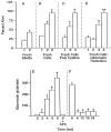
Toxicity is transferred by conditioned medium. Cell viability was measured after 24 hr in all cultures.A, Cells cultured for 8 d were exposed to 10 μm glutamate for 10 min, washed once, and returned to their original growth medium. After 11 hr either the cells were given fresh culture medium (2) or the medium was left undisturbed (1). B, In another experiment the medium was transferred to new cells of identical age in the absence (1) or presence (2) of 100 μm AP-5 or in the presence of AP-5 and 30 μm Ac-YVAD-cmk (3). C, Cells were exposed to growth-conditioned medium alone (1) or in the presence of 2 mm cystine (2) or in the presence of 2 mm cystine and 100 μm AP-5 (3). Because of the relative insolubility of cystine below pH 8, the experiments with cystine and all controls were performed at pH 8 by the reduction of incubator CO2.D, After 11 hr of glutamate exposure, growth-conditioned media were in some cases (2, 4) pretreated for 2 hr with GPT to reduce glutamate and then were transferred to fresh cells. 1, Growth-conditioned medium alone; 2, medium treated with GPT; 3, untreated medium plus AP-5; 4, glutamate-depleted medium plus AP-5. **Bar4 is significantly different from bar3,p < 0.01; n = 3.E, Concentration of glutamate in the growth-conditioned medium as a function of time after the addition of 10 μmglutamate to cultures. In one set of cultures 100 μm AP-5 was added before the addition of 10 μm glutamate (9 hr plus AP-5). F, Reduction of glutamate in the medium by GPT. GPT and cofactors were added to the growth-conditioned medium before application to the cells initially for 2 hr; then GPT was added repeatedly every 6 hr during the experiment to keep extracellular glutamate below 50 μm. *Significantly different from control (conditioned medium alone), p < 0.05;n = 3.

References

    1. Akazawa C, Shigemoto R, Bessho Y, Nakanishi S, Mizuno N. Differential expression of five N-methyl-d-aspartate receptor subunit mRNAs in the cerebellum of developing and adult rats. J Comp Neurol. 1994;347:150–160. - PubMed
    1. Ankacrona M, Dypbukt JM, Bonfoco E, Zhivotovsky B, Orrenius S, Lipton SA, Nicotera P. Glutamate-induced neuronal death: a succession of necrosis or apoptosis depending on mitochondrial function. Neuron. 1995;15:961–973. - PubMed
    1. Brewer GJ, Torricelli JR, Evege EK, Price PJ. Optimized survival of hippocampal neurons in B27-supplemented Neurobasal, a new serum-free medium combination. J Neurosci Res. 1993;35:567–576. - PubMed
    1. Carriedo SG, Yin HZ, Sensi SL, Weiss JH. Rapid Ca2+ entry through Ca2+-permeable AMPA/kainate channels triggers marked intracellular Ca2+ rises and consequent oxygen radical production. J Neurosci. 1998;18:7727–7738. - PMC - PubMed
    1. Choi DW. Ionic dependence of glutamate neurotoxicity in cortical cell culture. J Neurosci. 1987;7:369–379. - PMC - PubMed

Publication types

MeSH terms

LinkOut - more resources