. 2001 Oct 1;21(19):7551-60.
doi: 10.1523/JNEUROSCI.21-19-07551.2001.

Beta-amyloid induces neuronal apoptosis via a mechanism that involves the c-Jun N-terminal kinase pathway and the induction of Fas ligand

Affiliations

Beta-amyloid induces neuronal apoptosis via a mechanism that involves the c-Jun N-terminal kinase pathway and the induction of Fas ligand

Y Morishima et al. J Neurosci. .

Abstract

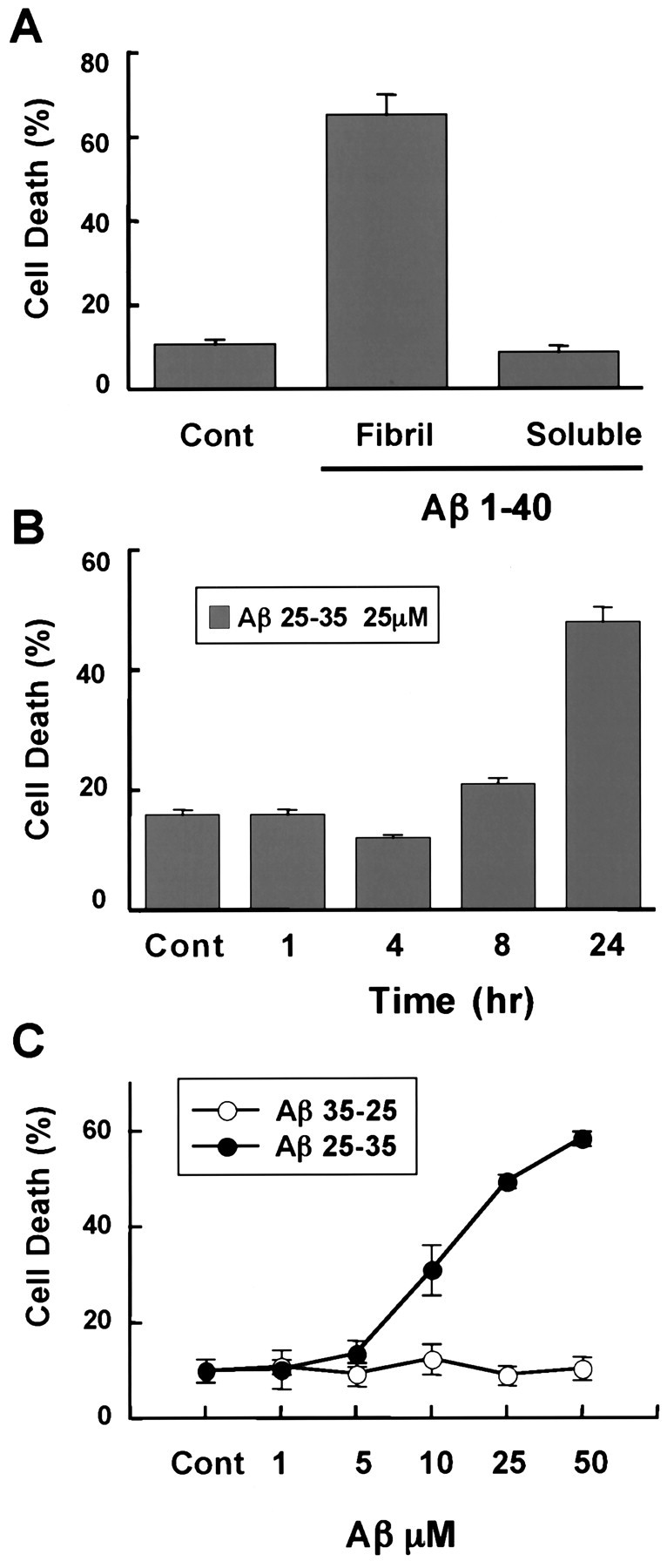
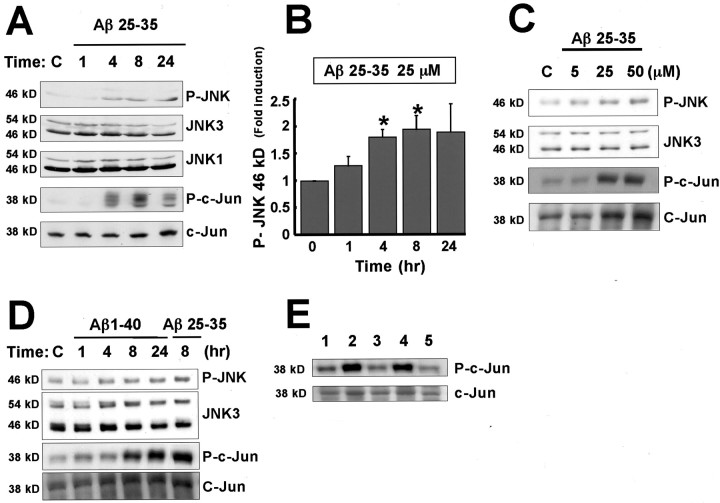
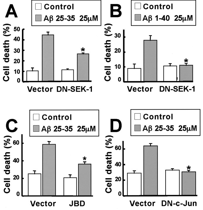
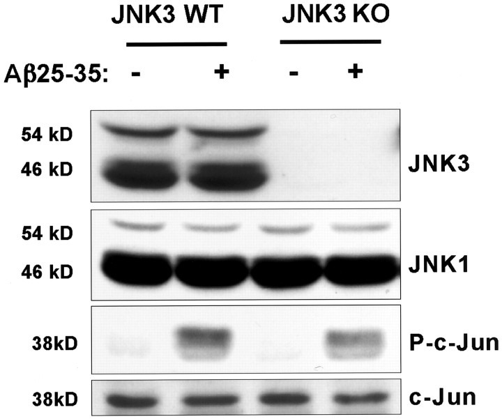
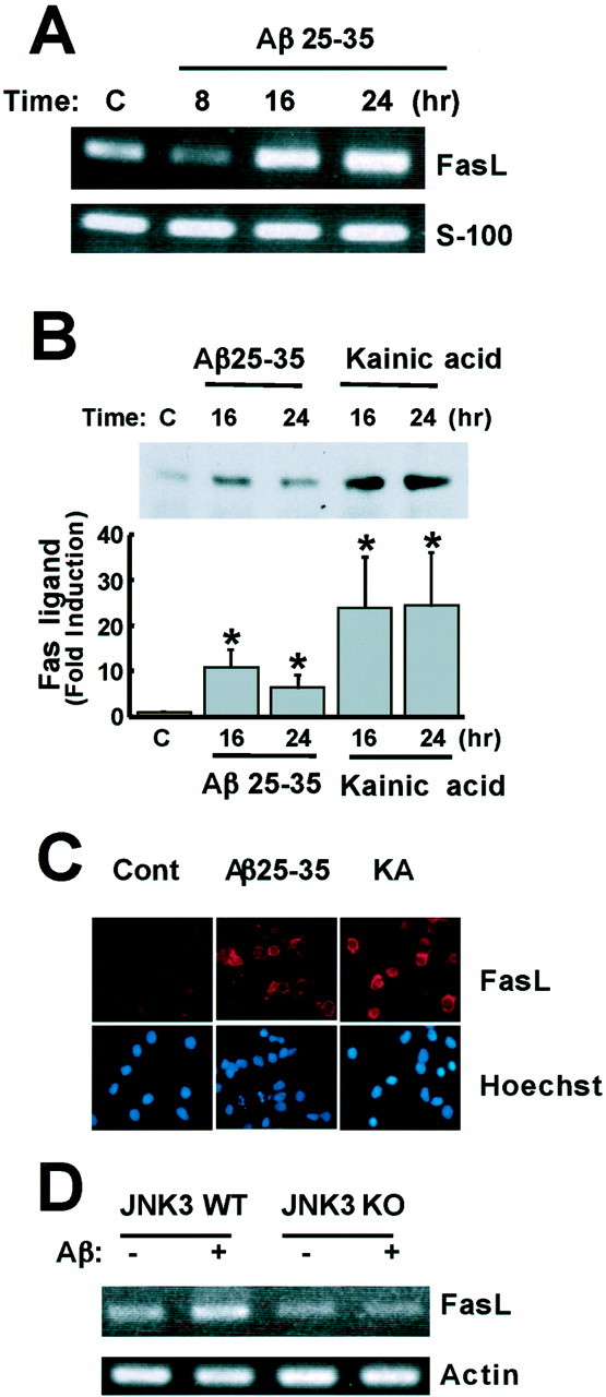
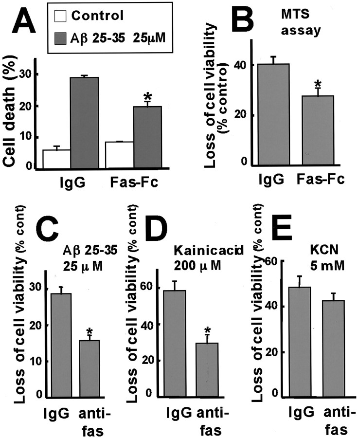
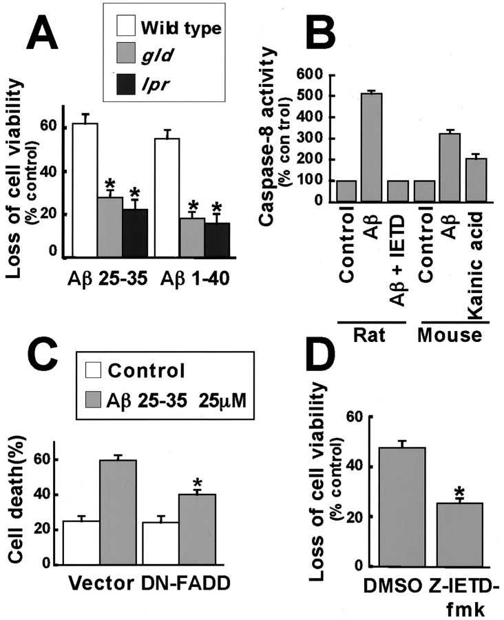
Elevated levels of beta-Amyloid (Abeta) are present in the brains of individuals with either the sporadic or familial form of Alzheimer's disease (AD), and the deposition of Abeta within the senile plaques that are a hallmark of AD is thought to be a primary cause of the cognitive dysfunction that occurs in AD. Recent evidence suggests that Abeta induces neuronal apoptosis in the brain and in primary neuronal cultures, and that this Abeta-induced neuronal death may be responsible in part for the cognitive decline found in AD patients. In this study we have characterized one mechanism by which Abeta induces neuronal death. We found that in cortical neurons exposed to Abeta, activated c-Jun N-terminal kinase (JNK) is required for the phosphorylation and activation of the c-Jun transcription factor, which in turn stimulates the transcription of several key target genes, including the death inducer Fas ligand. The binding of Fas ligand to its receptor Fas then induces a cascade of events that lead to caspase activation and ultimately cell death. By analyzing the effects of mutations in each of the components of the JNK-c-Jun-Fas ligand-Fas pathway, we demonstrate that this pathway plays a critical role in mediating Abeta-induced death of cultured neurons. These findings raise the possibility that the JNK pathway may also contribute to Abeta-dependent death in AD patients.


Figures

Fig. 1.
Fig. 1.
Aβ25–35 and Aβ1–40 promote neuronal cell death. A, Neurotoxicity of Aβ1–40. Rat primary cortical neurons were exposed to 50 μm fibrillar (Fibril) or soluble Aβ1–40 for 72 hr. Cells were fixed, nuclei were stained with Hoechst 33258, and apoptotic neurons were scored in a blinded manner as those cells that displayed condensed or fragmented nuclei. Experiments were performed at least three times, and data represent mean ± SEM of four wells from a representative experiment. B, Time course of Aβ25–35-induced neuronal apoptosis. Cultured neurons were treated with 25 μm Aβ25–35 for the indicated times, and apoptotic neurons were scored as described in A.C, Concentration-dependent neurotoxicity of Aβ25–35 and control peptide, Aβ35–25. Cortical neurons were exposed to Aβ25–35 or Aβ35-25 at various concentrations for 24 hr. Apoptotic neurons were scored as described in A.Cont, Control.
Fig. 2.
Fig. 2.
Aβ activates c-Jun and JNK in cortical neurons.A, Time course of Aβ-induced JNK and c-Jun phosphorylation. Rat cortical neurons were treated with 25 μm Aβ25–35 for the indicated number of hours. Whole-cell extracts were resolved by SDS-PAGE and immunoblotted with the antibodies directed against phospho-JNK or phospho-c-Jun (Ser-73). The total amount of JNK1, JNK3, and c-Jun protein was assessed using antibodies that recognize these proteins regardless of their phosphorylated state. B, Quantification of the phospho-JNK data (46 kDa isoform) from A. The densities of the bands were determined with a dual-wavelength Flying-spot scanner. *Statistical significance (p < 0.05) as assessed by the Wilcoxon test. C, Concentration-dependent activation of the JNK pathway by Aβ25–35. Cortical neurons were treated with Aβ25–35 at various concentrations for 8 hr. Western blotting analysis was done as described inA. D, Aβ1–40 activates the JNK pathway. Cortical neurons were incubated with 25 μmAβ1–40 or Aβ25–35 for the indicated times. Western blotting analysis was done as described in A. E, Aβ25–35 and fibril Aβ1–40 induce JNK phosphorylation in neurons. Cortical neurons were treated with PBS (lane 1), 25 μm Aβ25–35 (lane 2), 25 μm Aβ35–25 (lane 3), 25 μm fibrillar Aβ1–40 (lane 4), or 25 μm soluble Aβ1–40 (lane 5) for 8 hr. Western blotting analysis was performed using anti-phospho-specific c-Jun and anti-c-Jun as described in A.C, Control.
Fig. 3.
Fig. 3.
Dominant negative mutants of SEK-1 and c-Jun and the JBD of JIP-1 prevent Aβ-induced apoptosis in cortical neurons. Cortical neurons were cotransfected with 4 μg of an empty vector, DN-SEK-1, the JBD of JIP-1, or DN-c-Jun FlagΔ169 and 0.7 μg of CMV-LacZ and 0.3 μg of CMV-EGFP. Two or 1 d after the transfection, the neurons were treated with 25 μmAβ25–35 for 24 hr or 25 μm Aβ1–40 for 48 hr, respectively. The cells were fixed and immunostained with anti-β-Gal antibody. Nuclei were stained with Hoechst 33258. Transfected cells were identified as those that express green fluorescent protein. From the transfected cells, the apoptotic neurons were identified as those displaying condensed and fragmented nuclei. Data represent mean ± SEM of four wells from a typical experiment. *p < 0.05, significantly different from vector (ANOVA and Dunnett's test). Each experiment was repeated two or three times. A, Effect of DN-SEK-1 on Aβ25–35 toxicity. B, Effect of DN-SEK-1 on Aβ1–40 toxicity. C, Effect of the JBD of JIP-1 on Aβ25–35 toxicity. D, Effect of DN-c-Jun FlagΔ169 on Aβ25–35 toxicity.
Fig. 4.
Fig. 4.
Effect of Aβ on the expression levels of JNK1 and JNK3 and on the phosphorylation of c-Jun in cortical neurons from wild-type and JNK3−/− mice. Whole-cell extracts from wild-type and JNK3−/− cortical neurons treated with or without 25 μm Aβ25–35 (8 hr) were prepared, and equal amounts of proteins were resolved by SDS-PAGE. The levels of JNK1, JNK3, phosphorylated c-Jun, and total c-Jun were examined by immunoblotting. WT, Wild type;KO, knock-out.
Fig. 5.
Fig. 5.
JNK3-deficient neurons are resistant to Aβ-induced apoptosis. A, B, Cortical (A) and hippocampal (B) neurons were cultured from wild-type and JNK3−/−mice embryos and treated with 25 μm Aβ25–35 for 24 hr. Cells were fixed, and nuclei were stained with Hoechst 33258. Apoptotic neurons were scored in a blinded manner. Data represent mean ± SEM of four wells from a typical experiment. *p < 0.05, significantly different from wild type (ANOVA and Dunnett's test); n = 4. C, Wild-type and JNK3−/− cortical neurons were treated with 25 μm Aβ1–40 for 48 hr, and apoptotic neurons were counted as described in A. D, Wild-type and JNK3−/− cortical and hippocampal neurons were treated with 25 μm Aβ25–35 as described inA. Cell viability was assayed by metabolic integrity (MTS assay). Data represent mean ± SEM (n = 4). *p < 0.05, significantly different from wild type (Student's t test). KO, Knock-out; WT, wild type.
Fig. 6.
Fig. 6.
Aβ upregulates Fas ligand expression in cortical neurons. A, Aβ induction of Fas ligand (FasL) mRNA. Cortical neurons were treated with 25 μm Aβ25–35, and total RNA was extracted at the indicated times. Expression of Fas ligand and S100 mRNAs was examined by semiquantitative RT-PCR. B, Induction of Fas ligand protein level by Aβ and kainic acid. Cortical neurons were treated with 25 μm Aβ25–35 or 200 μm kainic acid, and total cell extracts were prepared at the indicated times. The level of Fas ligand was determined by immunoblotting using anti-Fas ligand antibodies and quantified using the Wilcoxon test (*p < 0.05). C, Immunostaining using anti-Fas ligand (FasL) antibody. Neurons were treated with Aβ25–35 or kainic acid (KA) for 16 hr and fixed. The cells were incubated with anti-Fas ligand antibody, followed by anti-mouse biotin-conjugated secondary antibody and then Cy3-labeled streptoavidin and Hoechst 33258. D, Cortical neurons derived from wild-type and JNK3−/− mice were treated with 25 μm Aβ25–35, and total RNA was extracted at 24 hr. Expression of Fas ligand and actin mRNAs was examined by semiquantitative RT-PCR. WT, Wild type;KO, knock-out.
Fig. 7.
Fig. 7.
Aβ induces apoptosis by a Fas ligand–Fas-dependent mechanism. A, B, Cortical neurons were pretreated with 15 μg/ml Fas–Fc or control IgG for 2 hr, and then 25 μm Aβ25–35 was added, and apoptosis was determined by scoring cells with condensed and fragmented nuclei (A) or using the MTS assay (B). *p < 0.05, significantly different from IgG (ANOVA and Dunnett's test or Student's t test). C–E, Cortical neurons were incubated with 2.5 μg/ml anti-Fas antibody or control IgG for 2 hr and then treated with 25 μm Aβ25–35 (C), 200 μm kainic acid (D), or 5 mm KCN (E). Cell death was assessed by the MTS assay. *p < 0.05, significantly different from IgG (Student's t test).
Fig. 8.
Fig. 8.
The Fas ligand–Fas pathway is required for Aβ-induced apoptosis. A, Cortical neurons were prepared from wild-type, gld, and lprmice embryos and treated with 25 μm Aβ25–35 for 24 hr or 25 μm Aβ1–40 for 72 hr. Cell viability was assessed by the MTS assay. Data represent mean ± SEM (n = 5). *p < 0.05, significantly different from wild type (ANOVA and Dunnett's test).B, Neurons from rat and mouse were treated with 25 μm Aβ25–35 and the caspase-8 inhibitor, Ζ-IETD-fmk, or with 200 μm kainic acid. Caspase-8 activity was assessed by a fluorometric assay kit. Data represent mean ± SEM (n = 6 for rats; n = 4 for mice). C, Cortical neurons were cotransfected with 4 μg of an empty vector or DN-FADD and 0.7 μg of CMV-LacZ and 0.3 μg of CMV-EGFP. After 2 d, the cells were treated with 25 μm Aβ25–35 for 24 hr. The cells were fixed and immunostained with anti-β-Gal antibody. Nuclei were stained with Hoechst 33258. Transfected cells were identified as those that expressed green fluorescent protein, and of these cells, apoptotic neurons were scored as those that display condensed and fragmented nuclei. Data represent mean ± SEM of four wells from a typical experiment. D, Neurons were pretreated with DMSO or 100 μm caspase 8 inhibitor Ζ-IETD-fmk for 1 hr, and then 25 μm Aβ25–35 was added, and apoptosis was determined by the MTS assay. Data represent mean ± SEM of seven wells from a typical experiment. *p < 0.05, significantly different from vector or control (ANOVA and Dunnett's test or Student's t test).

References

    1. Anderson AJ, Cummings BJ, Cotman CW. Increased immunoreactivity for Jun- and Fos-related proteins in Alzheimer's disease. Exp Neurol. 1994;125:286–295. - PubMed
    1. Anderson AJ, Su JH, Cotman CW. DNA damage and apoptosis in Alzheimer's disease: colocalization with c-Jun immunoreactivity, relationship to brain area, and effect of postmortem delay. J Neurosci. 1996;16:1710–1719. - PMC - PubMed
    1. Behrens A, Sibilis M, Wagner EF. Amino-terminal phosphorylation of c-Jun regulates stress-induced apoptosis and cellular proliferation. Nat Genet. 1999;21:326–329. - PubMed
    1. Brunet A, Bonni A, Zigmond MJ, Lin MZ, Juo P, Hu LS, Anderson MJ, Arden KC, Blenis J, Greenberg ME. Akt promotes cell survival by phospholylating and inhibiting a forkhead transcription factor. Cell. 1999;96:857–868. - PubMed
    1. Chang BS, Minn AJ, Muchmore SW, Fesik SW, Thompson CB. Identification of a novel regulatory domain in Bcl-xL and Bcl-2. EMBO J. 1997;16:968–977. - PMC - PubMed

Publication types

MeSH terms