. 2001 Oct 1;21(19):7608-19.
doi: 10.1523/JNEUROSCI.21-19-07608.2001.

Neurons lacking huntingtin differentially colonize brain and survive in chimeric mice

Affiliations

Neurons lacking huntingtin differentially colonize brain and survive in chimeric mice

A Reiner et al. J Neurosci. .

Abstract

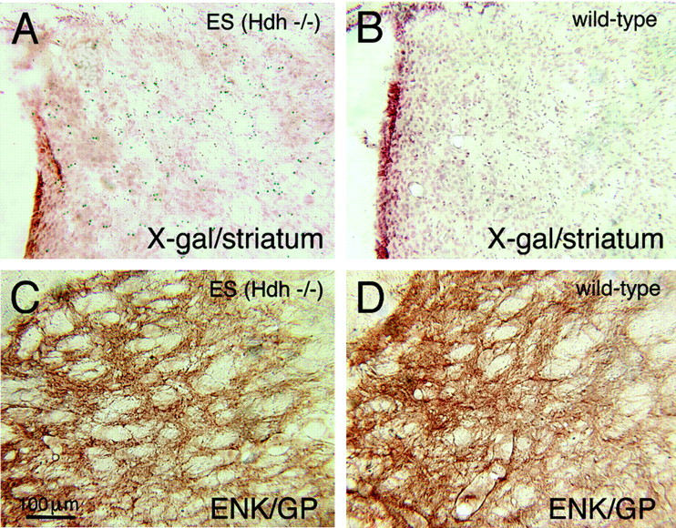
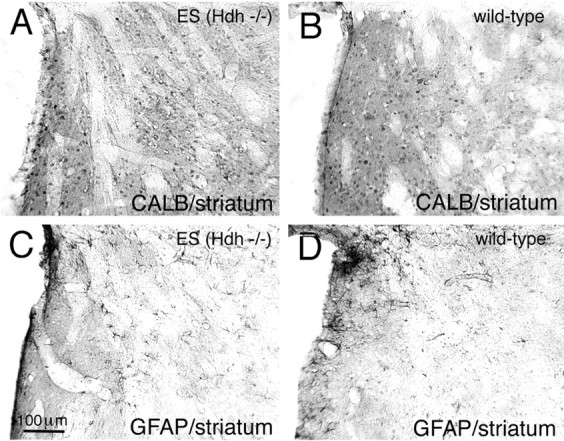
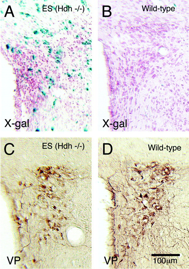
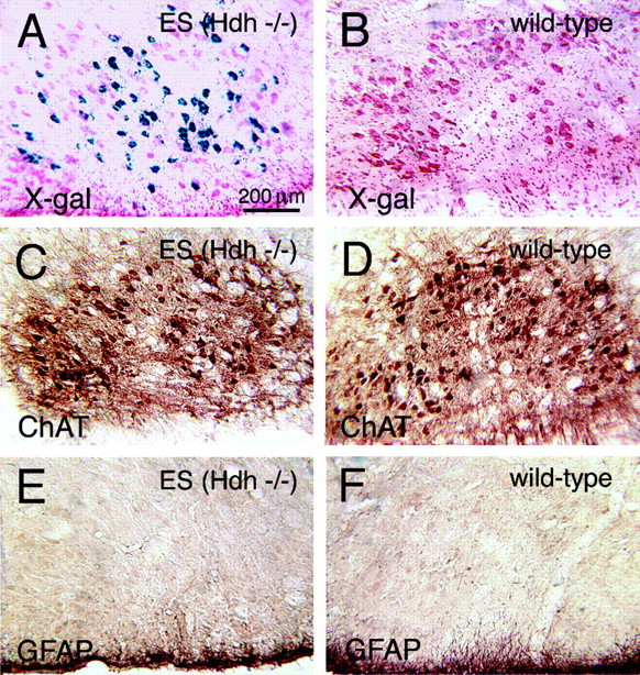
To determine whether neurons lacking huntingtin can participate in development and survive in postnatal brain, we used two approaches in an effort to create mice consisting of wild-type cells and cells without huntingtin. In one approach, chimeras were created by aggregating the 4-8 cell embryos from matings of Hdh (+/-) mice with wild-type 4-8 cell embryos. No chimeric offspring that possessed homozygous Hdh (-/-) cells were obtained thereby, although statistical considerations suggest that such chimeras should have been created. By contrast, Hdh (-/-) ES cells injected into blastocysts yielded offspring that were born and in adulthood were found to have Hdh (-/-) neurons throughout brain. The Hdh (-/-) cells were, however, 5-10 times more common in hypothalamus, midbrain, and hindbrain than in telencephalon and thalamus. Chimeric animals tended to be smaller than wild-type littermates, and chimeric mice rich in Hdh (-/-) cells tended to show motor abnormalities. Nonetheless, no brain malformations or pathologies were evident. The apparent failure of aggregation chimeras possessing Hdh (-/-) cells to survive to birth is likely attributable to the previously demonstrated critical role of huntingtin in extraembryonic membranes. That Hdh (-/-) cells in chimeric mice created by blastocyst injection are under-represented in adult telencephalon and thalamus implies a role for huntingtin in the development of these regions, whereas the neurological dysfunction in brains enriched in Hdh (-/-) cells suggests a role for huntingtin in adult brain. Nonetheless, the lengthy survival of Hdh (-/-) cells in adult chimeric mice indicates that individual neurons in many brain regions do not require huntingtin to participate in normal brain development and to survive.


Figures

Fig. 1.
Fig. 1.
Image of a sagittal section from one of the chimeric mice created by blastocyst injection of Hdh−/− ES cells, stained to reveal the location of the X-gal-positive Hdh−/− ES cell progeny that have colonized the brain. The tissue was counterstained with neutral red. This animal (ES13) was among those chimeric mice possessing Hdh−/− cells that were killed before 1 year of age because of signs of morbidity. The green–blue X-gal labeling shows that Hdh −/− cells are found throughout brain but are most abundant in hippocampus, preoptic area, hypothalamus, midbrain, and hindbrain.
Fig. 2.
Fig. 2.
Image of CA2 in a transverse section from one of the chimeric mice created by blastocyst injection of Hdh−/− ES cells, stained to reveal the localization of huntingtin-containing wild-type cells (visualized by DAB immunolabeling; A) and X-gal-positive ES cell progeny that have colonized the hippocampus (B). The X-gal-labeled tissue also was counterstained with neutral red. This animal was among those that lived up to 1 year of age with no signs of morbidity. The green–blue X-gal labeling shows thatHdh −/− CA2 pyramidal cells in regions lacking in X-gal labeling are immunostained for huntingtin (arrow indicates a large cluster of huntingtin-labeled neurons). By contrast, regions of CA2 richly labeled for X-gal (arrowhead) are poor in or devoid of huntingtin immunolabeling.
Fig. 3.
Fig. 3.
High-magnification images showing the distribution and abundance of Hdh −/− cells in several brain regions in the same chimeric mouse created by blastocyst injection of Hdh −/− ES cells, as shown in Figure 1. The green–blue X-gal labeling shows thatHdh −/− cells are scarce in striatum (A) and highly abundant in the preoptic region (B) and dorsal pons (C). The image presented in Dshows that the vast majority of the neurons in the facial nucleus in this case was Hdh −/−. The section from which these images were taken had been counterstained lightly with neutral red.
Fig. 4.
Fig. 4.
High-magnification images of transverse sections through dorsomedial striatum showing the distribution and abundance ofHdh −/− cells (as visualized by X-gal labeling, followed by a light neutral red counterstain) in the striatum of a chimeric mouse created by blastocyst injection ofHdh −/− ES cells. Shown is the striatum of a chimeric mouse, who displayed no ill health up to 1 year of age (A), compared with the striatum of a wild-type mouse in which no Hdh −/−cells are present (B). C,D, The presence of Hdh−/−cells in the striatum of the chimeric mouse shown in Ahas not produced any evident abnormality in the enkephalinergic striatal output fibers (ENK; visualized by DAB immunolabeling) within the ipsilateral globus pallidus (GP) of the chimeric mouse (C). Medial is to the left and dorsal to thetop in all images.
Fig. 5.
Fig. 5.
High-magnification images of transverse sections through dorsomedial striatum of the same chimeric and wild-type animals as shown in Figure 4. The presence of Hdh−/− cells in the striatum of the chimeric mouse has not produced any evident abnormality in the labeling of calbindergic striatal perikarya (A, B) or any evidence of neuropathology in the striatum, as shown by the absence of any upregulation of glial fibrillary acid protein (GFAP) in the striatum (C,D). Medial is to the left and dorsal to the top in all images.
Fig. 6.
Fig. 6.
High-magnification images of transverse sections through the paraventricular nucleus of the hypothalamus showing the distribution and abundance of Hdh−/− cells in striatum of a chimeric mouse created by blastocyst injection of Hdh−/− ES cells (ES8). Shown is the striatum of a chimeric mouse, who displayed no ill health up to 1 year of age (A), compared with the paraventricular nucleus of a wild-type mouse in which no Hdh−/− cells are present (B). The Hdh−/− cells in A are visualized by X-gal labeling, and neuronal cytoarchitecture in bothA and B is visualized by neutral red counterstaining. C, D, The presence ofHdh −/− cells in the paraventricular nucleus of the chimeric mouse shown in Ahas not produced any evident abnormality in the vasopressinergic (VP) neurons (visualized by DAB immunolabeling) of the ipsilateral paraventricular nucleus of the chimeric mouse. Medial is to the left and dorsal to the top in all images.
Fig. 7.
Fig. 7.
High-magnification images of transverse sections through pons showing the distribution and abundance of Hdh−/− cells in the facial nucleus in a chimeric mouse created by blastocyst injection of Hdh−/− ES cells. Shown is the facial nucleus in a chimeric mouse, who displayed no ill health up to 1 year of age (A), compared with facial nucleus of a wild-type mouse in which no Hdh −/− cells are present (B). The Hdh−/− cells in A are visualized by X-gal labeling, and neuronal cytoarchitecture in bothA and B is visualized by neutral red counterstaining. C, D, The presence ofHdh −/− cells in the facial nucleus of the chimeric mouse shown in A has not produced any evident abnormality in the facial motoneurons (visualized by DAB immunolabeling for choline acetyltransferase) within the ipsilateral facial nucleus of the chimeric mouse. High-magnification images of transverse sections through facial nucleus of the same chimeric and wild-type animals as shown in A and Breveal that the presence of Hdh −/−cells in the facial nucleus of the chimeric mouse has not produced any upregulation of GFAP in the facial nucleus (E,F). Medial is to the left and dorsal to the top in all images.

References

    1. Albin RL, Tagle DA. Genetics and molecular biology of Huntington's disease. Trends Neurosci. 1995;18:11–14. - PubMed
    1. Albin RL, Reiner A, Anderson KD, Penney JB, Young AB. Striatal and nigral neuron subpopulations in rigid Huntington's disease: implications for the functional anatomy of chorea and rigidity-akinesia. Ann Neurol. 1990a;27:357–365. - PubMed
    1. Albin RL, Young AB, Penney JB, Handelin B, Balfour R, Anderson KD, Markel DS, Tourtellotte WW, Reiner A. Abnormalities of striatal projection neurons and N-methyl-d-aspartate receptors in presymptomatic Huntington's disease. N Engl J Med. 1990b;332:1293–1298. - PubMed
    1. Albin RL, Reiner A, Anderson KD, Dure LS, IV, Handelin B, Balfour R, Whetsell WO, Jr, Penney JB, Young AB. Preferential loss of striato-external pallidal projection neurons in presymptomatic Huntington's disease. Ann Neurol. 1992;31:425–430. - PubMed
    1. Ambrose CM, Duyao MP, Barnes G, Bates GP, Lin CS, Srinidhi J, Baxendale S, Hummerich H, Lehrach H, Atherr M, Wasmuth J, Buckler A, Church D, Housman D, Berks M, Micklem G, Durbin R, Dodge A, Read A, Gusella J, MacDonald ME. Evidence against simple inactivation due to an expanded CAG repeat. Somat Cell Mol Genet. 1994;20:27–38. - PubMed

Publication types

MeSH terms