. 2001 Oct 1;21(19):7630-41.
doi: 10.1523/JNEUROSCI.21-19-07630.2001.

Delayed inner ear maturation and neuronal loss in postnatal Igf-1-deficient mice

Affiliations

Delayed inner ear maturation and neuronal loss in postnatal Igf-1-deficient mice

G Camarero et al. J Neurosci. .

Abstract

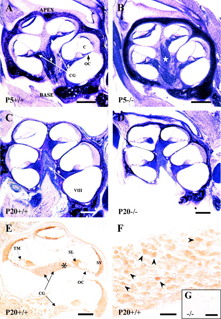
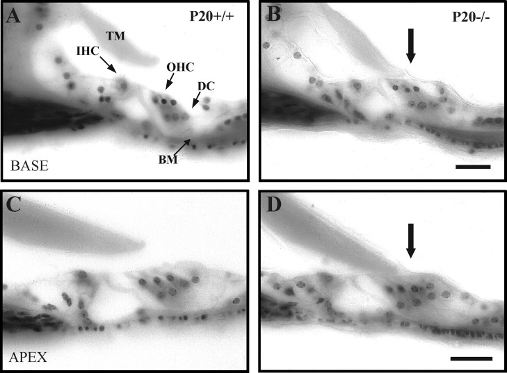
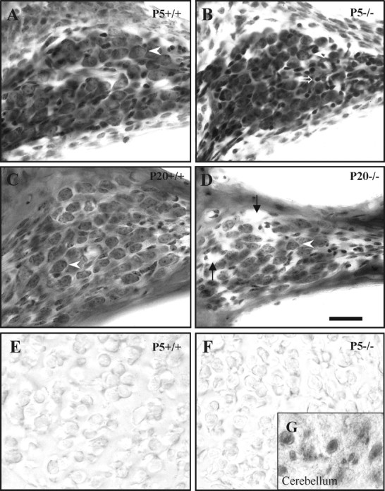
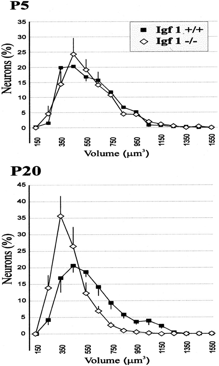
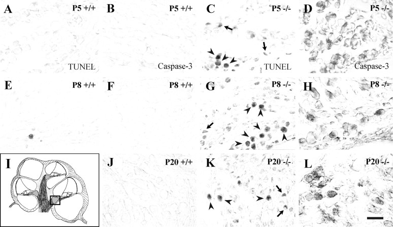
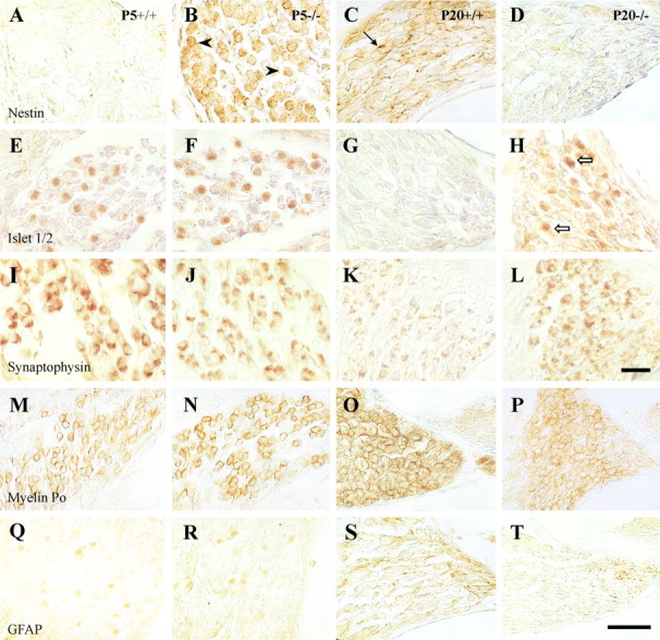
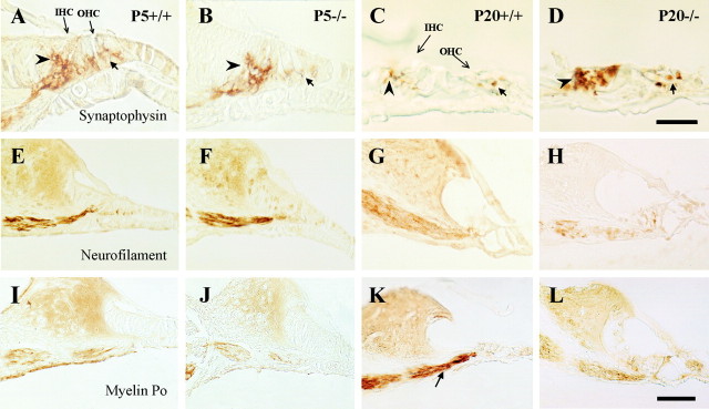
Insulin-like growth factor-1 (IGF-1) has been shown to play a key role during embryonic and postnatal development of the CNS, but its effect on a sensory organ has not been studied in vivo. Therefore, we examined cochlear growth, differentiation, and maturation in Igf-1 gene knock-out mice at postnatal days 5 (P5), P8, and P20 by using stereological methods and immunohistochemistry. Mutant mice showed reduction in size of the cochlea and cochlear ganglion. An immature tectorial membrane and a significant decrease in the number and size of auditory neurons were also evident at P20. IGF-1-deficient cochlear neurons showed increased caspase-3-mediated apoptosis, along with aberrant expression of the early neural markers nestin and Islet 1/2. Cochlear ganglion and fibers innervating the sensory cells of the organ of Corti presented decreased levels of neurofilament and myelin P(0) in P20 mouse mutants. In addition, an abnormal synaptophysin expression in the somata of cochlear ganglion neurons and sensory hair cells suggested the persistence of an immature pattern of synapses distribution in the organ of Corti of these animals. These results demonstrate that lack of IGF-1 in mice severely affects postnatal survival, differentiation, and maturation of the cochlear ganglion cells and causes abnormal innervation of the sensory cells in the organ of Corti.


Figures

Fig. 1.
Fig. 1.
Cochlear anatomy of the mouse genotypes analyzed and IGF-1 expression. Nissl staining of mid-modiolar celloidin sections of the cochlea at P5 (A, B) or P20 (C, D) in Igf-1+/+ (A, C) and Igf-1−/− (B, D) mice. An increased thickness of the otic capsule cartilage and a dispersion of the fibers in the auditory branch of the eighth cranial nerve of Igf-1−/− at P5 (star) were observed in all animals studied. Note the reduction in size ofIgf-1−/− P20 cochlea compared with the Igf-1+/+ P20 controls. IGF-1 immunohistochemical expression in the cochlea (E) and cochlear ganglia (F) of P20Igf-1+/+ mice.Asterisk in E indicates the area shown inF; arrowheads in F point to the subpopulation of IGF-1-positive neurons. Theinset shows a magnification of the cochlear ganglion ofIgf-1−/− P20 animals (G). C, Cochlear duct;O, otic capsule; OC, organ of Corti;CG, cochlear ganglion; VIII, eighth cranial nerve; TM, tectorial membrane;SL, spiral limbus; SV, stria vascularis. Scale bars: A–D, 650 μm; E, 600 μm;F, 55 μm; G, 10 μm.
Fig. 2.
Fig. 2.
Altered morphology of the OC tectorial membrane in Igf-1−/− P20 mice. Nissl staining of celloidin-embedded cross sections of the OC of P20Igf-1+/+ (A, C) andIgf-1−/− (B, D) animals. A and B show basal turns, whereas C and D show apical turns of the cochlea. Physical attachment of the tectorial membrane to the hair cells was noticed in all sections of P20Igf-1−/− (B, D, arrows). BM, Basilar membrane;DC, Deiters' cells; IHC, inner hair cells; OHC, outer hair cells; TM,tectorial membrane. Scale bars, 30 μm.
Fig. 3.
Fig. 3.
Altered cochlear ganglion morphology inIgf-1−/− mice. Nissl staining of the ganglion in the cochlear basal turn inIgf-1+/+ (A, C) andIgf-1−/− (B, D) mice at P5 (A, B) and P20 (C, D). At P5, the cochlear ganglion and its neurons show similar morphology in both genotypes (arrowheads), except thatIgf-1−/− mouse also show a subtype of abnormally small, strongly chromaffinic cells (open arrow). At P20, ganglion cells are noticeably reduced both in size and number in Igf-1−/− mice. This reduction, despite the presence of enlarged intercellular spaces (arrows), leads to a considerable decrease in the ganglion cross-sectional area. E and Fshow negative PCNA expression in the cochlear ganglion cells of P5Igf-1+/+ (E) and Igf-1−/−(F) mice. The inset(G) shows a positive control of PCNA-positive cerebellum cells from the same section of P20Igf-1−/− mouse shown inF. Scale bar, 30 μm.
Fig. 4.
Fig. 4.
Different perikaryal volume distributions of cochlear ganglion neurons in Igf-1+/+ andIgf-1−/− mice. Linear plots of data obtained by application of stereological methods (see Materials and Methods) are represented for P5 (top) and P20 (bottom) mice. Error bars represent the SD among animals for each volume class. Whereas the distribution is monomodal in all cases, note the leftward displacement of the curve in the mutant mice at P20, with a marked decrease of cells in the largest size classes. See Table2 for statistical analysis.
Fig. 5.
Fig. 5.
Apoptotic cell death in the cochlear ganglion ofIgf-1−/− mice. TUNEL labeling (A, C, E, G, K) and detection of activated caspase-3 expression (B, D, F, H, J, L) were performed on paraffin sections from normal (Igf-1+/+) and mutant (Igf-1−/−) mice at postnatal days 5, 8, and 20. The area analyzed is shown in a schematic drawing of the cochlea in which the square indicates basal turn cochlear ganglia (I). Note the increase in apoptotic nuclei (C, G, K) and intense activated caspase-3 immunostaining (D, H, L) in the mutant mice.Arrowheads point to apoptotic neurons, whereasarrows point to dying glial cells. The sections correspond to basal turns of the cochlea. Scale bar, 30 μm.
Fig. 6.
Fig. 6.
Delayed differentiation of postnatal cochlear ganglion in Igf-1−/− mice. Immunohistochemical analysis of paraffin sections of the cochlear ganglion at midmodiolar levels using nestin (A–D), Islet-1/2 (E–H), synaptophysin (I–L), myelin P0(M–P), and GFAP (Q–T) antibodies. Left panels (A,E, I, M,Q) correspond to P5 Igf-1+/+ samples,middle-left panels (B, F, J, N,R) to P5 Igf-1−/−,middle-right panels (C, G, K, O,S) are from P20Igf-1+/+, and right panels (D, H, L, P, T) from P20 Igf-1−/− mice.E–H correspond to basal turns of the cochlear ganglion, and the remaining sections in the figure are from apical turns. Scale bars: A–L, 40 μm; M–T, 30 μm.
Fig. 7.
Fig. 7.
Differential expression of neural and glial markers in the innervation of the organ of Corti. Immunohistochemical staining of midmodiolar paraffin sections of basal turns of the cochlea with synaptophysin (A–D), neurofilament 200 kDa (E–H), and myelin P0(I–L) antibodies. Left panels(A, E, I) correspond to P5Igf-1+/+ samples, middle-left panels (B, F, J) to P5Igf-1−/−, middle-right panels (C, G, K) to P20Igf-1+/+, and right panels (D, H, L) to P20Igf-1−/− mice. At P5 (A, B), synaptophysin immunoreactivity appears diffuse and displays a “cup-like” shape surrounding the IHCs (arrowheads) and OHCs (arrows). At P20 (C, D), the synaptophysin staining pattern is better defined at the base of IHC and nerve fibers in the Igf-1+/+ controls than in Igf-1−/− mutants. NF-200K and myelin P0 immunostainings do not evidence major differences at P5 but are clearly less intense in P20Igf-1−/− mice (H, L) than in Igf-1+/+ controls (G, K, arrow). Scale bars: A–D, 20 μm;E–L, 30 μm.

References

    1. Adesanya OO, Zhou J, Samathanam C, Powell-Braxton L, Bondy CA. Insulin-like growth factor 1 is required for G2 progression in the estradiol-induced mitotic cycle. Proc Natl Acad Sci USA. 1999;96:3287–3291. - PMC - PubMed
    1. Avendaño C, Dykes RW. Quantitative analysis of anatomical changes in the cuneate nucleus following forelimb denervation: a stereological morphometric study in adult cats. J Comp Neurol. 1996;370:491–500. - PubMed
    1. Baker J, Liu JP, Robertson EJ, Efstratiadis A. Role of insulin-like growth factors in embryonic and postnatal growth. Cell. 1993;75:73–82. - PubMed
    1. Baker J, Hardy MP, Zhou J, Bondy C, Lupu F, Bellve AR, Efstratiadis A. Effects of an Igf1 gene null mutation on mouse reproduction. Mol Endocrinol. 1996;10:903–918. - PubMed
    1. Beck KD, Powell-Braxton L, Widmer HR, Valverde J, Hefti F. Igf1 gene disruption results in reduced brain size, CNS hypomyelination, and loss of hippocampal granule and striatal parvalbumin-containing neurons. Neuron. 1995;14:717–730. - PubMed

Publication types

MeSH terms

Substances