. 2001 Nov;69(5):961-8.
doi: 10.1086/323764. Epub 2001 Sep 20.

Syndromic short stature in patients with a germline mutation in the LIM homeobox LHX4

Affiliations

Syndromic short stature in patients with a germline mutation in the LIM homeobox LHX4

K Machinis et al. Am J Hum Genet. 2001 Nov.

Abstract

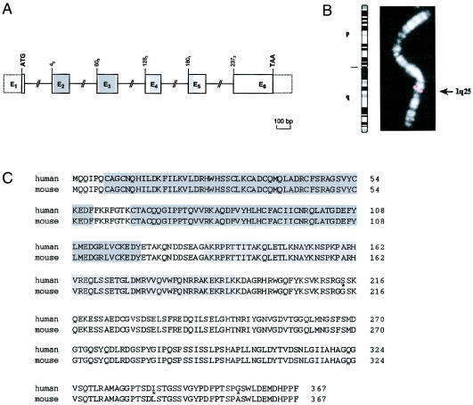
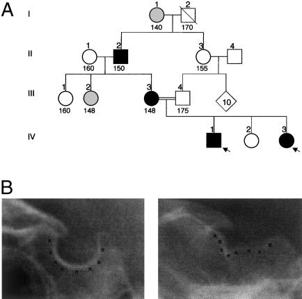
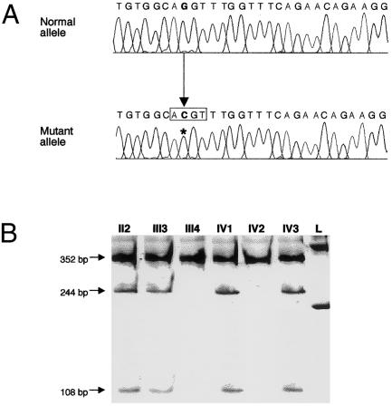
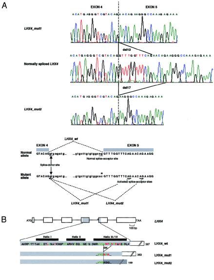
Studies of genetically engineered flies and mice have revealed the role that orthologs of the human LIM homeobox LHX4 have in the control of motor-neuron-identity assignment and in pituitary development. Remarkably, these mouse strains, which bear a targeted modification of Lhx4 in the heterozygous state, are asymptomatic, whereas homozygous animals die shortly after birth. Nevertheless, we have isolated the human LHX4 gene, as well as the corresponding cDNA sequence, to test whether it could be involved in developmental defects of the human pituitary region. LHX4, which encodes a protein 99% identical to its murine counterpart, consists of six coding exons and spans >45 kb of the q25 region of chromosome 1. We report a family with an LHX4 germline splice-site mutation that results in a disease phenotype characterized by short stature and by pituitary and hindbrain (i.e., cerebellar) defects in combination with abnormalities of the sella turcica of the central skull base. This intronic mutation, which segregates in a dominant and fully penetrant manner over three generations, abolishes normal LHX4 splicing and activates two exonic cryptic splice sites, thereby predicting two different proteins deleted in their homeodomain sequence. These findings, which elucidate the molecular basis of a complex Mendelian disorder, reveal the fundamental pleiotropic role played by a single factor that tightly coordinates brain development and skull shaping during head morphogenesis.


Figures

Figure  1
Figure 1
Genomic structure, chromosomal localization, and predicted amino acid sequence of human LHX4. A, Intron-exon organization of human LHX4. The six exons (E1–E6) encoding LHX4 are indicated by boxes with either solid or dashed borders, depicting translated or untranslated sequences, respectively. Sequences encoding the two LIM domains and the homeodomain are shaded dark gray and light gray, respectively. The locations of the ATG initiation codon and of the TAA stop codon are shown. The number of the first codon in each exon is indicated; exons beginning with the first, second, or third base of a codon are indicated by the subscripts “1,” “2,” or “3,” respectively. Exons are drawn to scale. B, Chromosomal mapping of human LHX4 by FISH. The fine localization of LHX4 to 1q25 is indicated by the black arrow (right). An ideogram of chromosome 1 is given (left). C, Comparison of deduced amino acid sequences of human and mouse LHX4. Different residues are indicated by an asterisk. The two LIM domains and the homeodomain are shaded dark gray and light gray, respectively.
Figure  2
Figure 2
Pedigree and phenotypic features of kindred presenting with brain and skull developmental abnormalities. A, Pedigree of family. Black symbols denote affected individuals, gray symbols denote individuals with uncertain phenotypic status, and white symbols denote unaffected individuals; circles denote females, and squares denote males; a slash through a symbol denotes a deceased individual. Although individuals I1 and III2 exhibit short stature—as in the patients’ phenotype—their phenotypic status remains uncertain, because no further clinical and biological information was available. Heights (in cm) are indicated under the corresponding symbols. Probands are indicated by arrows. B, Radiographs of sella turcica region. The shapes of sella turcica from a normal individual (left) and from patient IV3 (right) are highlighted by black Xs.
Figure  3
Figure 3
Identification of LHX4 molecular defect. A, Nucleotide sequences of normal (top) and mutant (bottom) LHX4 alleles, as determined after cloning of the LHX4 PCR products into TOPO-XL cloning vectors (Invitrogen). The G→C substitution (boldface; position indicated by asterisk) identified in the mutant allele creates a recognition sequence for the MaeII restriction enzyme (box). B, Segregation analysis of splice-acceptor–site mutation in the family. The presence of the mutation in the genomic DNA of six family members was assessed by means of MaeII digestion of PCR products generated by primers bracketing the splice site. This assay confirmed that all affected members tested (lanes II2, III3, IV1, and IV3) have one mutant (244 bp and 108 bp) and one normal (352 bp) allele, whereas the unaffected individuals (III4 and IV2) have only normal (352 bp) alleles. The size marker (lane L) is the “Smart-ladder” (Eurogentec).
Figure  4
Figure 4
Consequences of LHX4 intronic mutation, at RNA level and protein level. A, Effect that mutation has on splicing of LHX4 primary transcripts. Top, Sequence electropherograms of normal and mutant LHX4 transcripts. The mutant allele generates two populations of transcripts—one with a deletion of 12 nucleotides (LHX4_mut1) and another with a deletion of 17 nucleotides (LHX4_mut2), both located within the coding sequence of exon 5. Bottom, Schematic representation of in vitro splicing of normal and mutant LHX4. The location of the G→C substitution is indicated by an asterisk. The invariant splice-donor site and the three splice-acceptor sites (i.e., the site normally used, as well as the two cryptic splice sites that are activated in the mutated allele) are underlined. B, Evolutionary conservation of amino acid residues involved in LHX4-homeodomain deletions generated by mutant allele. Top, Schematic presentation of genomic structure of LHX4. Exons are drawn to scale and are depicted by rectangles. (Introns are not drawn to scale.) Sequences from exons 4 and 5 encode the homeodomain (shaded). The splice-acceptor–site mutation (indicated by an asterisk) is located upstream from exon 5. Bottom, Normal and mutant LHX4-homeodomain amino acid sequences. The amino acid sequence of normal LHX4 (LHX4_wt) is shown against a light-gray background. Helices are denoted by black boxes. In the consensus sequence, the seven positions occupied by amino acids that are invariant in >95% of the homeodomain proteins thus far identified are indicated by red letters. Highly conserved residues (i.e., at the most, three different amino acids at these locations in all homeodomain proteins thus far identified) are indicated by green letters (Burglin, T. R.). The mutant allele generates two populations of transcripts (encoding mutant LHX4 proteins)—one maintaining the normal reading frame and another changing the reading frame and resulting in the creation of a premature termination codon within exon 5. The four amino acids, which are deleted when either of the two cryptic splice-acceptor sites is used, are indicated on the normal LHX4-homeodomain sequence (del). The LHX4-homeodomain products (LHX4_mut1 and LHX4_mut2) resulting from the mutation are drawn to scale, in proportion to the length of the normal homeodomain. Part of the nonsense sequence, which results from the frameshift introduced by the activation of the splice-acceptor site located 17 nucleotides downstream of the normal one, is shown in italic against a dark-gray background. Numbers indicate the total length of predicted amino acid sequences.

References

Electronic-Database Information

    1. Burglin, T. R., http://www.biosci.ki.se/groups/tbu/homeo/consensus.gif (for information relevant to homeobox proteins and their classification and evolution)
    1. GenBank, http://www.ncbi.nlm.nih.gov/Genbank/ (for mouse Lhx4 cDNA sequence [accession number AF135415] and for human LHX4 cDNA sequence [accession number AF282899])
    1. GeneMap'99, http://www.ncbi.nlm.nih.gov/genemap99/ (for in silico mapping of human ESTs)
    1. Online Mendelian Inheritance in Man (OMIM), http://www.ncbi.nlm.nih.gov/Omim/ (for Chiari type I malformation [MIM 118420] and Lhx4 [MIM 602146])

References

    1. Burset M, Seledtsov IA, Solovyev VV (2001) SpliceDB: database of canonical and non-canonical mammalian splice sites. Nucleic Acids Res 29:255–259 - PMC - PubMed
    1. Couly G, Le Douarin NM (1988) The fate map of the cephalic neural primordium at the presomitic to the 3-somite stage in the avian embryo. Development 103:101–113 - PubMed
    1. Dattani MT, Martinez-Barbera JP, Thomas PQ, Brickman JM, Gupta R, Martensson IL, Toresson H, Fox M, Wales JK, Hindmarsh PC, Krauss S, Beddington RS, Robinson IC (1998) Mutations in the homeobox gene HESX1/Hesx1 associated with septo-optic dysplasia in human and mouse. Nat Genet 19:125–133 - PubMed
    1. Dubois PM, El Amraoui A, Héritier AG (1997) Development and differentiation of pituitary cells. Microsc Res Tech 39:98–113 - PubMed
    1. Eagleson GW, Harris WA (1990) Mapping of the presumptive brain regions in the neural plate of Xenopus laevis. J Neurobiol 21:427–440 - PubMed

Publication types

MeSH terms

Associated data