

Review
doi: 10.1136/bjo.85.10.1252.
A hypothesis to explain ganglion cell death caused by vascular insults at the optic nerve head: possible implication for the treatment of glaucoma
Affiliations
- PMID: 11567974
- PMCID: PMC1723727
- DOI: 10.1136/bjo.85.10.1252
Item in Clipboard
Review
A hypothesis to explain ganglion cell death caused by vascular insults at the optic nerve head: possible implication for the treatment of glaucoma
Br J Ophthalmol.
2001 Oct.
No abstract available
Figures
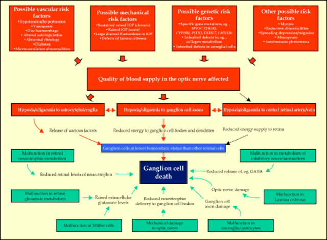
Possible causes of ganglion cell death in glaucoma. It is suggested that activation of sufficient risk factors causes the quality of blood supply in the optic nerve head to be affected. As a result, the nutritional supply to the optic nerve head is slowly compromised (oligaemia/hypoxia) particularly affecting astrocytes, microglia, and ganglion cell axons. Even the central retinal artery/vein may become slightly affected. Such insults eventually lead to the death of ganglion cells as depicted and detailed in Figures 2 and 3. One may also envisage other modes of stimulating ganglion cell death in glaucoma (lighter blue boxes) where the vascular system does not have a direct role. Some of these possibilities may also be linked to certain risk factors, but these are not indicated.
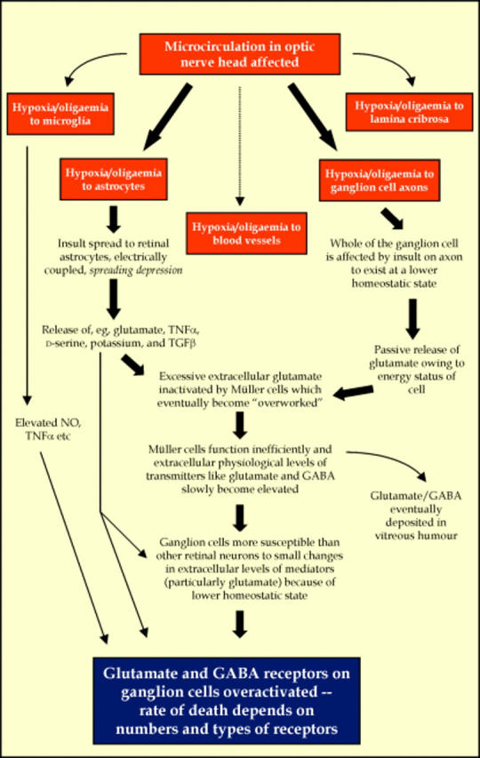
A hypothesis to explain ganglion cell death in glaucoma. Various components in the optic nerve head may be affected by oligaemia/hypoxia as a result of an alteration in the microcirculation (see Fig 1). While the ganglion cell axon may be affected in the initial stages of the insult, the whole of the cell will eventually suffer (exist at a lower homeostatic state) with glutamate particularly being "non-physiologically" released into the extracellular space (Fig 3). Astrocytes and microglial cells are also likely to release a variety of substances into the extracellular space after an undefined duration of insult. Some of these substances may have "protective" properties while others will have adverse effects on neurons. Moreover, increased levels of glutamate in the extracellular space are potentially toxic to many retinal cells. Müller cells will as a consequence become particularly active in an attempt to maintain physiological levels of extracellular neurotransmitters. However, the excessive demands placed on Müller cells will eventually lead to them becoming inefficient. This will result in a slow but gradual rise in the level of glutamate and other neurotransmitters (for example, GABA) in the extracellular space. The ganglion cells, being at a lower homeostatic status than other retinal cell types will potentially, therefore, be more susceptible to this extracellular rise of neurotransmitters. It is proposed that at a certain point, glutamate will overexcite ganglion cells to initiate a dying process. It is also hypothesised that the variability in the death rate of individual ganglion cells will depend on the degree of this overexcitement, which is dependent in part on the number of excitatory and inhibitory receptors associated with the neuron (and also upon a rise in the extracellular levels of neurotransmitters). Activation of inhibitory GABA receptors, for example, will hyperpolarise the cell and this will tend to counteract the overexcitation.
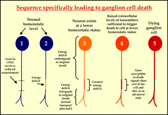
Proposed stages for ganglion cell death in glaucoma (see Figure 2 for details).
References
Publication types
MeSH terms
LinkOut - more resources
Full Text Sources
Other Literature Sources
Medical