Comparative Study
. 2001 Jul;111(7):1285-9.
doi: 10.1097/00005537-200107000-00027.

Motility-related proteins as markers for head and neck squamous cell cancer

Affiliations
Comparative Study

Motility-related proteins as markers for head and neck squamous cell cancer

M T Abraham et al. Laryngoscope. 2001 Jul.

Abstract

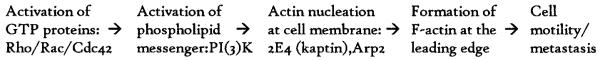
Hypothesis: Increased cell motility is a hallmark of cancer cells. Proteins involved in cell motility may be used as molecular markers to characterize the malignant potential of tumors.

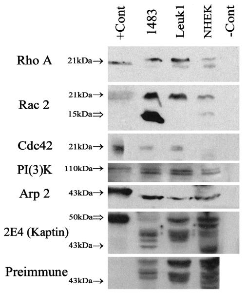
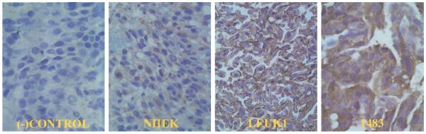
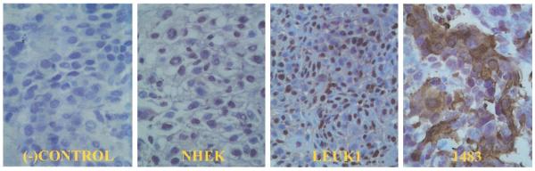
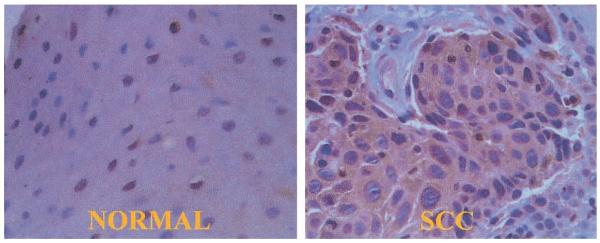
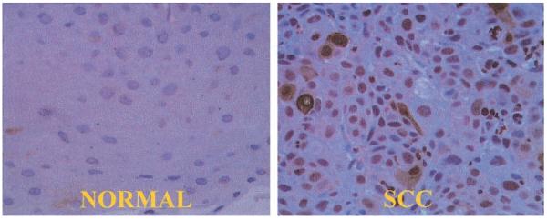
Methods: Molecular biology and immunohistochemistry techniques were used to investigate the expression of a selected panel of motility-related proteins (Rho A, Rac 2, Cdc42, PI3K, 2E4, and Arp2) in normal, premalignant, and squamous cell cancer cell lines of human head and neck origin. To assess the clinical potential of these proteins as molecular markers for cancer, immunohistochemistry was performed on paraffin-fixed head and neck cancer specimens (n = 15).

Results: All six motility-associated proteins were overexpressed in the premalignant and squamous cell cancer cell lines relative to normal keratinocytes. Immunohistochemistry with Rho A and Rac 2 showed increased staining in areas of cancer but not in normal tissue.

Conclusion: Proteins involved in cell motility can be used as markers for head and neck squamous cell carcinoma. The head and neck cell lines used in this study may be used as a model to further investigate cell motility. Molecular markers of motility could have a significant impact on the diagnosis and staging of cancers originating from differentiated non-motile cells.


Figures

Fig. 1
Fig. 1
Proposed simplified cascade of molecular events necessary for cell motility and tumor cell metastasis. Unknown intermediary signaling molecules may be necessary.
Fig. 2
Fig. 2
Rho A, Rac 2, Cdc42, and Arp 2 Western blots of whole cell lysates separated by 12% SDS-PAGE, and PI(3)K, 2E4 (kaptin), and rabbit Preimmune Western blots separated by 10% SDS-PAGE. Bands are present at the expected molecular weight of each protein (solid arrows: Rho A—21 kDA, Rac 2—21 kDa, Cdc42—21 kDa, PI(3)K—110 kDa, Arp 2—43 kDa, and 2E4—43 kDa). These bands are present in the positive control lane (except for 2E4, see explanation below), absent in the negative control lane, and increased in intensity in the malignant 1483 squamous cell cancer and premalignant Leuk1 dysplastic lanes relative to the normal NHEK keratinocytes lane on each blot, respectively. On the Rac 2 blot, there is a second intensely positive band (open arrow) at 15 kDa in the malignant 1483 lane, which appears to be absent or at a much lower concentration in the premalignant Leuk1 and normal NHEK lanes. This band may represent a breakdown product of Rac 2 and is addressed further in the Discussion. On the 2E4 blot, a band is present at 50 kDa in the positive control lane, the expected molecular weight of the recombinant bacterially expressed 2E4 protein (open arrow). As expected, this band is present in the malignant 1483 lane at 43 kDa, the expected molecular weight of native 2E4 (solid arrow). This band is absent in the negative control lane, premalignant Leuk1 lane, and normal NHEK lane on the 2E4 blot, and is absent in all lanes on the Preimmune blot (there is some overlap of nonspecific bands in the NHEK lane). Rho A +Cont = HeLa whole cell lysate; Rac 2 +Cont = HL-60 whole cell lysate; Cdc42 +Cont = HeLa whole cell lysate; PI(3)K +Cont = COLO 320 DM whole cell lysate; Arp 2 +Cont = purified recombinant bacterially expressed Arp 2; 2E4 +Cont = purified recombinant bacterially expressed 2E4; 1483 = malignant squamous cell cancer cell line; Leuk1 = premalignant dysplastic cell line; NHEK = normal human epidermal keratinocytes; −Cont = malignant squamous cell cancer (1483) whole cell lysate without secondary antibody.
Fig. 3
Fig. 3
Compact cell block immunohistochemistry of cell lines with Rho A. Rho A is a cytoplasmic protein. As expected, there is no Rho A staining in the negative control panel. The expression of Rho A is sequentially increased in the premalignant Leuk1 and malignant 1483 cell lines relative to the normal NHEK cell line. (−)Control = 1483 cell line without primary antibody; NHEK = normal human epidermal keratinocytes; Leuk1 = premalignant dysplastic cell line; 1483 = malignant squamous cell cancer cell line (40× magnification).
Fig. 4
Fig. 4
Compact cell block immunohistochemistry of cell lines with Rac 2. As expected, there is no Rac 2 staining in the negative control panel. Rac 2 is a cytoplasmic protein. Cytoplastic staining of Rac 2 is present only in a subset of malignant 1483 cells. Sequentially increased nuclear staining is evident in the premalignant Leuk1 and malignant 1483 cell lines relative to the normal NHEK cell line. Refer to discussion for explanation. (−)Control = 1483 cell line without primary antibody; NHEK = normal human epidermal keratinocytes; Leuk1 = premalignant dysplastic cell line; 1483 = malignant squamous cell cancer cell line (40× magnification).
Fig. 5
Fig. 5
Rho A immunohistochemistry of a moderately differentiated squamous cell cancer of the tongue surgical specimen. In the normal panel, it is evident that there is minimal staining of the normal tissue adjacent to the cancer. The nest of squamous cell cancer (SCC) cells is clearly stained by the Rho A antibody in the SCC panel. Rho A is a cytoplasmic protein (40× magnification).
Fig. 6
Fig. 6
Rac 2 immunohistochemistry of a moderately differentiated squamous cell cancer of the tongue surgical specimen. In the normal panel, there is minimal staining of the normal tissue adjacent to the cancer. In the SCC (squamous cell cancer) panel, there is both cytoplasmic and nuclear staining of squamous cell cancer cells. Note that not all cancer cells express cytoplasmic Rac 2. Refer to Discussion for explanation (40× magnification).

References

    1. Liotta L. Mechanisms of cancer invasion and metastasis. In: DeVita VT, Hellman A, Rosenberg S, editors. Progress in Oncology. vol 1. Lippincott; New York: 1985. pp. 28–42.
    1. Cunningham CC. Actin structural proteins in cell motility. Cancer Metastasis Rev. 1992;11:69–77. - PubMed
    1. Bearer EL. Role of actin polymerization in cell locomotion: molecules and models. Am J Respir Cell Mol Biol. 1993;8:582–591. - PMC - PubMed
    1. Ridley AJ. The small GTP-binding rho regulates the assembly of focal adhesions and actin stress fibers in response to growth factors. Cell. 1992;70:389–399. - PubMed
    1. Ridley AJ, Paterson HF, Johnston CL, Diekmann D, Hall A. The small GTP-binding protein Rac regulates growth factor-induced membrane ruffling. Cell. 1992;70:401–410. - PubMed

Publication types

MeSH terms