

Lack of correlation between Ki-67 labelling index and tumor size of anterior pituitary adenomas
- PMID: 11570981
- PMCID: PMC56633
- DOI: 10.1186/1471-2407-1-12
Lack of correlation between Ki-67 labelling index and tumor size of anterior pituitary adenomas
Abstract
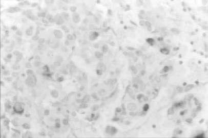
Aims and background: The Ki-67 is a nuclear antigen detected by the monoclonal antibody MIB-1 and its Labeling Index (LI) is considered a marker of normal and abnormal cell proliferation. Pituitary adenomas are generally well differentiated neoplasms, even if in about one third of cases they are invasive of surrounding tissues. The aim of this study is to evaluate the correlation between Ki-67 labelling index and tumor size of pituitary adenomas extimated by means CT and MRI and confirmed at operation.
Methods: Using the monoclonal antibody MIB-1, we evaluated the expression of Ki-67 in 121 anterior pituitary adenomas consecutively operated on in a 48-month period.
Results: In relation to neuroradiological (CT and MRI) and surgically verified tumor size, we identified 24 microadenomas, 27 intrasellar macroadenomas, 34 intra-suprasellar macroadenomas, and 36 intra-supra-parasellar macroadenomas. The adenomas were non-infiltrating (76 cases) and infiltrating (45 cases) adenomas. The wall of the cavernous sinus (CS) was infiltrated in 18 cases. Forty-eight adenomas were non-functioning and 73 functioning. The overall mean +/- SD Ki-67 LI was 2.72 +/- 2.49% (median 1.6). It was 2.59 +/- 1.81 in microadenomas, 2.63 +/- 3.45 in intrasellar macroadenomas, 1.91 +/- 2.11 in intra-suprasellar macroadenomas, and 3.29 +/- 5.45 in intra-supra-parasellar macroadenomas (p = 0.27). It was 3.73 +/- 5.13% in infiltrating and 2.03 +/- 2.41% in non-infiltrating adenomas (p = 0.02), and 5.61 +/- 7.19% in CS-infiltrating versus 2.09 +/- 2.37% in CS-non-infiltrating adenomas (p = 0.0005).
Conclusions: Our preliminary results seem to exclude significative correlations between Ki-67 LI and tumor size of anterior pituitary adenomas, even if this index can be considered a useful marker in the determination of the infiltrative behaviour of these tumors.
Figures
References
-
- Asano K, Kubo O, Tajika Y, Huang MC, Takakura K. The relationship between cell proliferation and secretory activity in pituitary adenomas. A review of 63 cases. No To Shinkei. 1996;48:543–549. - PubMed
-
- Atkin SL, Green VL, Hipkin LJ, Landolt AM, Foy PM, Jeffreys RV, et al. A comparison of proliferation indices in human anterior pituitary adenomas using formalin-fixed tissue and in vitro cell culture. J Neurosurg. 1997;87:85–88. - PubMed
-
- Brown DC, Gatter KC. Monoclonal antibody Ki-67: its use in histopathology. Histopathology. 1990;17:489–503. - PubMed
-
- Buchfelder M, Fahlbusch R, Adams EF, Kiesewetter F, Thierauf P. Proliferation parameters for pituitary adenomas. Acta Neurochir Suppl (Wien) 1996;65:18–21. - PubMed
-
- Daita G, Yonemasu Y. Dural invasion and proliferative potential of pituitary adenomas. Neurol Med Chir. 1996;36:211–214. - PubMed
MeSH terms
Substances
LinkOut - more resources
Full Text Sources
Medical
Molecular Biology Databases
