

Resolution of viable and membrane-compromised bacteria in freshwater and marine waters based on analytical flow cytometry and nucleic acid double staining
- PMID: 11571170
- PMCID: PMC93217
- DOI: 10.1128/AEM.67.10.4662-4670.2001
Resolution of viable and membrane-compromised bacteria in freshwater and marine waters based on analytical flow cytometry and nucleic acid double staining
Abstract
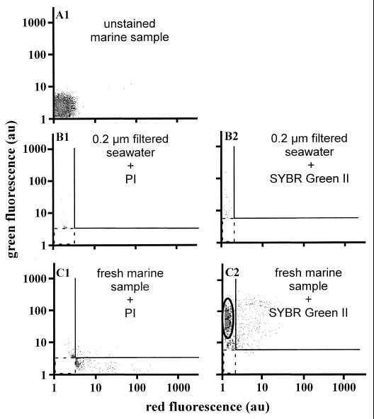
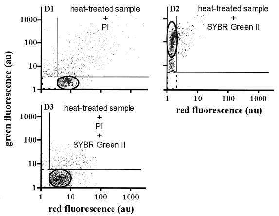
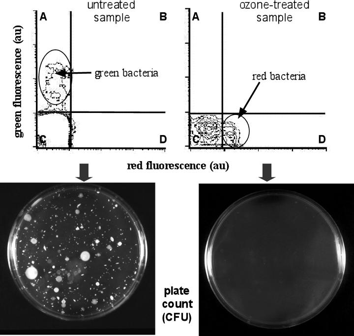
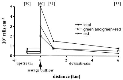
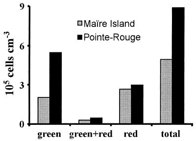
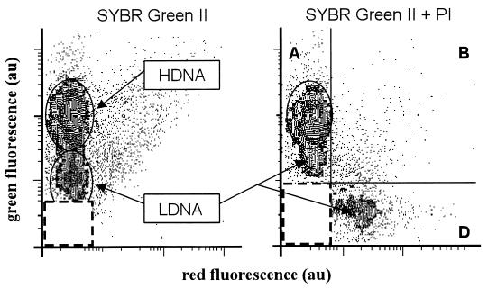
The membrane integrity of a cell is a well-accepted criterion for characterizing viable (active or inactive) cells and distinguishing them from damaged and membrane-compromised cells. This information is of major importance in studies of the function of microbial assemblages in natural environments, in order to assign bulk activities measured by various methods to the very active cells that are effectively responsible for the observations. To achieve this task for bacteria in freshwater and marine waters, we propose a nucleic acid double-staining assay based on analytical flow cytometry, which allows us to distinguish viable from damaged and membrane-compromised bacteria and to sort out noise and detritus. This method is derived from the work of S. Barbesti et al. (Cytometry 40:214-218, 2000) which was conducted on cultured bacteria. The principle of this approach is to use simultaneously a permeant (SYBR Green; Molecular Probes) and an impermeant (propidium iodide) probe and to take advantage of the energy transfer which occurs between them when both probes are staining nucleic acids. A full quenching of the permeant probe fluorescence by the impermeant probe will point to cells with a compromised membrane, a partial quenching will indicate cells with a slightly damaged membrane, and a lack of quenching will characterize intact membrane cells identified as viable. In the present study, this approach has been adapted to bacteria in freshwater and marine waters of the Mediterranean region. It is fast and easy to use and shows that a large fraction of bacteria with low DNA content can be composed of viable cells. Admittedly, limitations stem from the unknown behavior of unidentified species present in natural environments which may depart from the established permeability properties with respect to the fluorescing dyes.
Figures

References
-
- Alonso M C, Rodriguez V, Rodriguez J, Borrego J J. Role of ciliates, flagellates, and bacteriophages on the mortality of marine bacteria and on dissolved-DNA concentration in laboratory experimental systems. J Exp Mar Biol Ecol. 2000;244:239–252.
-
- Arnosti C, Repeta D J, Blough N V. Rapid bacterial degradation of polysaccharides in anoxic marine systems. Geochim Cosmochim Acta. 1994;58:2639–2652.
-
- Barbesti S, Citterio S, Labra M, Baroni M D, Neri M G, Sgorbati S. Two- and three-color fluorescence flow cytometric analysis of immunoidentified viable bacteria. Cytometry. 2000;40:214–218. - PubMed
-
- Barer M R, Harwood C R. Bacterial viability and culturability. Adv Microb Physiol. 1999;41:94–137. - PubMed
-
- Bianchi M. Nouvelles approches d'étude des réseaux microbiens. Ann Limnol. 1998;34:465–473.
Publication types
MeSH terms
Substances
LinkOut - more resources
Full Text Sources
