

Infection of Acanthamoeba polyphaga with Simkania negevensis and S. negevensis survival within amoebal cysts
- PMID: 11571186
- PMCID: PMC93233
- DOI: 10.1128/AEM.67.10.4789-4795.2001
Infection of Acanthamoeba polyphaga with Simkania negevensis and S. negevensis survival within amoebal cysts
Abstract
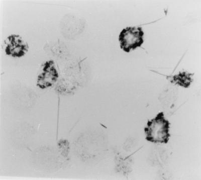
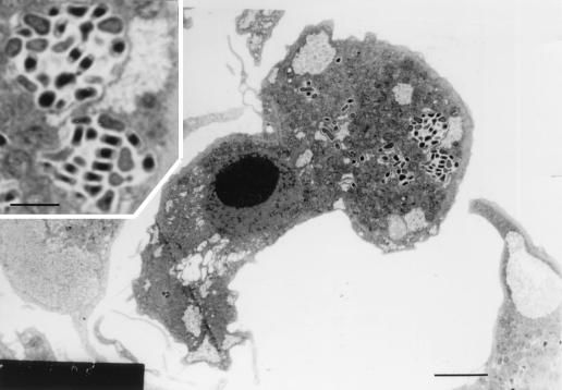
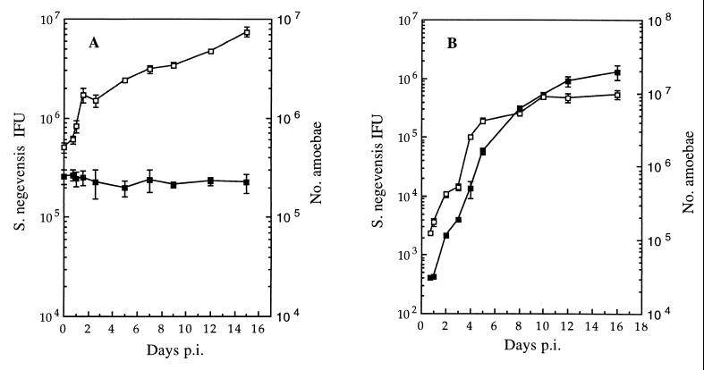
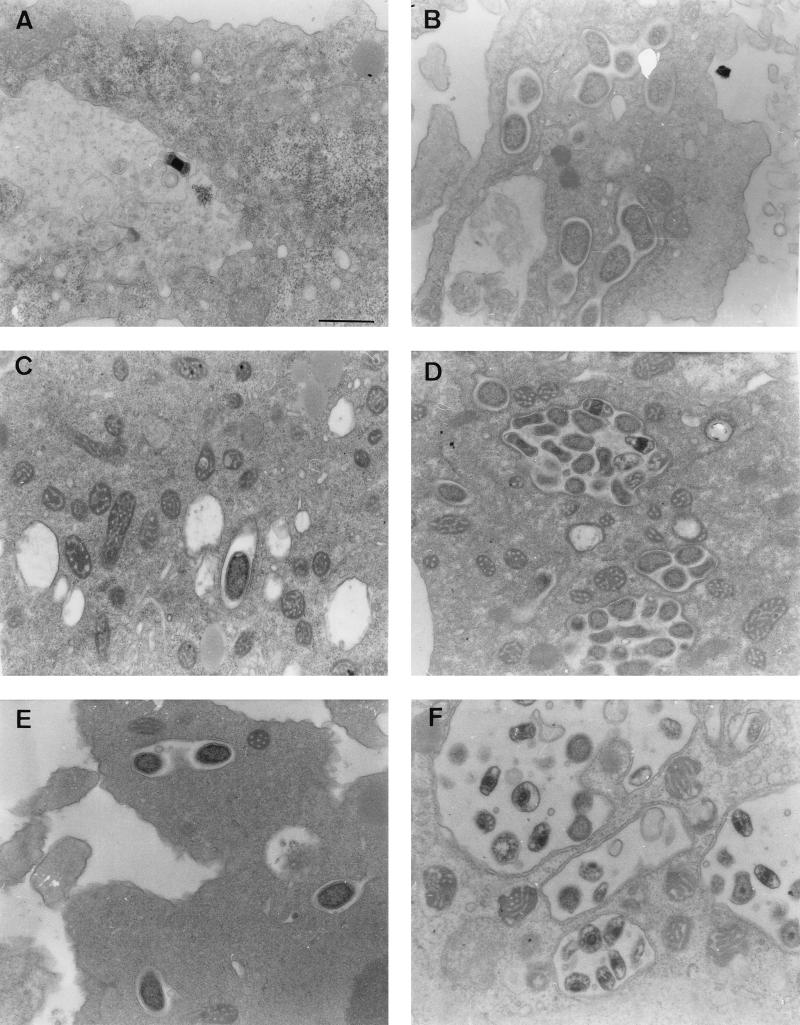
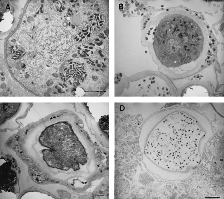
Simkania negevensis, a novel microorganism belonging to the family Simkaniaceae in the order Chlamydiales, has an intracellular developmental cycle during which two morphological entities, elementary bodies (EB) and reticulate bodies (RB), are seen by electron microscopy. Rates of seropositivity to the organism are high in certain population groups, and S. negevensis has been associated with respiratory illness in humans. This study reports for the first time the ability of S. negevensis to survive and grow inside Acanthamoeba polyphaga in addition to its known ability to grow in cell cultures of human or simian origin. Infectivity of S. negevensis and growth in amoebae were monitored by immunoperoxidase assays. Long-term persistence and exponential growth of S. negevensis in amoebal trophozoites were demonstrated by infectivity assays and by electron microscopy. EB and dividing RB of S. negevensis were observed within inclusion bodies inside A. polyphaga. When S. negevensis-infected A. polyphaga amoebae were exposed to adverse conditions resulting in encystation of the amoebae, several possible outcomes were observed: cysts containing both normal amoebic cytoplasm and S. negevensis; cysts in which S. negevensis cells were relegated to the space between cyst walls; and cysts containing S. negevensis, but apparently lacking amoebal cytoplasm. S. negevensis within dried amoebal cysts was capable of long-term survival. The possibility that amoebae may have a role in natural transmission of S. negevensis needs to be investigated.
Figures
References
-
- Armstrong M. The pathogenesis of human Acanthamoeba infection. Infect Dis Rev. 2000;2:65–73.
-
- Biberfeld P. Cytological studies on blood lymphocytes activated by phytohaemagglutinin in vitro. Acta Pathol Microbiol Scand. 1971;223:7–8. - PubMed
-
- Birtles R J, Rowbotham T J, Storey C, Marrie T J, Raoult D. Chlamydia-like obligate parasite of free-living amoebae. Lancet. 1997;349:925–930. - PubMed
Publication types
MeSH terms
Substances
LinkOut - more resources
Full Text Sources
